Tubulin protein (97% pure): porcine brain

Tubulin protein (97% pure): porcine brain
$0.00

Product Uses Include

  • Economical alternative to T240 in primary screens for tubulin ligand drugs
  • High-throughput tubulin polymerization or depolymerization screens
  • Production of microtubule substrates for HTS motor assays

Material
Tubulin protein has been isolated from porcine brain. The final product contains approximately 97% tubulin and 3% Microtubule Associated Proteins (MAPs). This version of porcine tubulin is an economical alternative to our highly purified tubulin (Cat. # T240) for anti-tubulin ligand drug discovery. It has been formulated to be compatible with 96 or 384-well format polymerization assays such as in Biochem™ kits BK004P or BK011P .


Purity
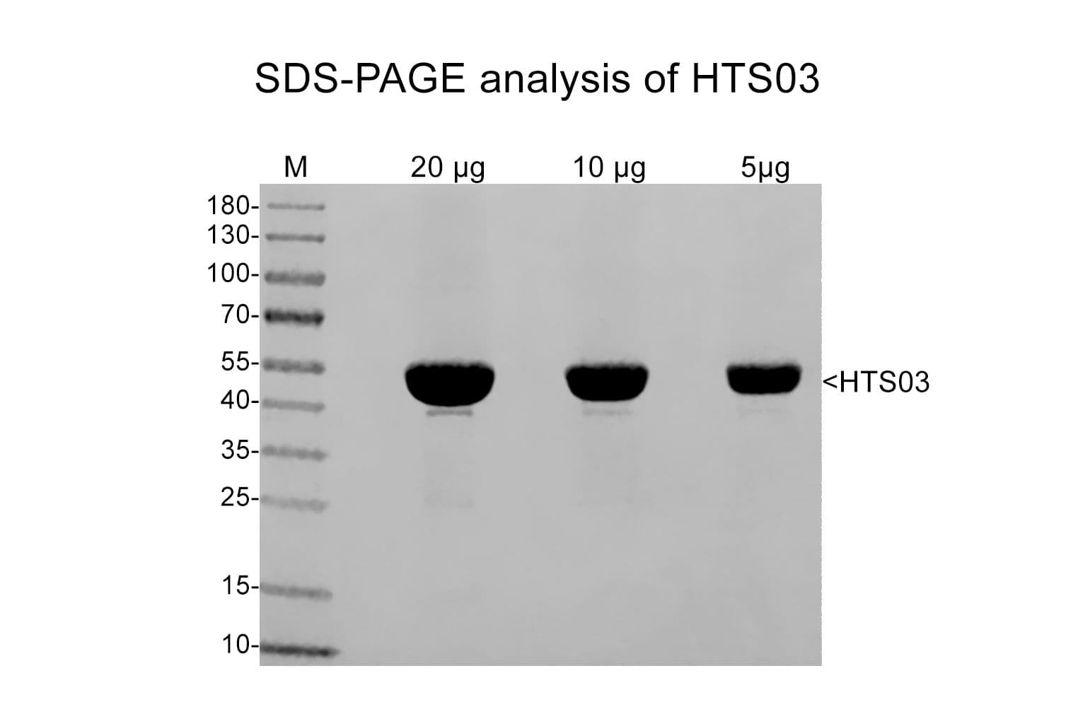
Protein purity is determined by scanning densitometry of Coomassie Blue stained protein on a 12% SDS-PAGE gel. HTS02 contains 97% tubulin (MW. 55 kDa) and 3% MAPs (MW 35-280 kDa).

hts02

Figure 1: A 100 µg sample of HTS03 protein was separated by electrophoresis on a 12% SDS-PAGE gel and stained with Coomassie Blue. Protein quantitation was performed using the Precision Red Protein Assay Reagent (Cat.# ADV02).

Biological Activity
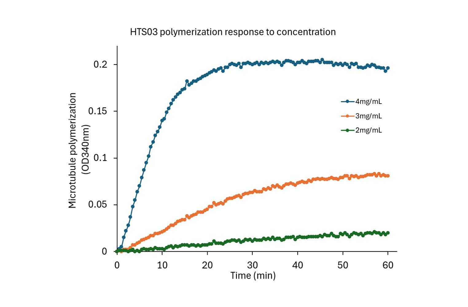
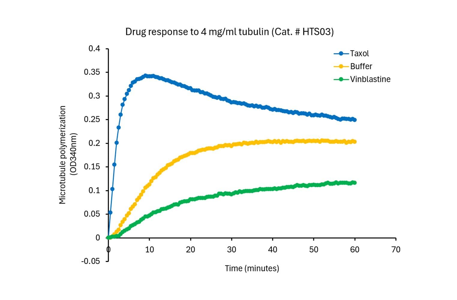
An ASSAY UNIT is defined as the amount of HTS03 protein needed to achieve a tubulin polymerization signal of OD340 of 0.1 - 0.15 in 30 minutes at 37°C in G-PEM buffer. The assay volume is 100 µl and assumes a spectrophotometer pathlength of 0.5 cm. The protein concentration for HTS02 in this assay is approximately 4 mg/ml. The biological activity of HTS03 is assessed by a tubulin polymerization assay. One ASSAY UNIT of tubulin is used for each polymeriztion assay. An OD340 of 0.10 - 0.15 is required to pass quality control. Tubulin polymerization must also be responsive to polymerization enhancers (paclitaxel) and inhibitors (nocodazole) at 5 µM drug concentration.

CDS01res

Figure 2: Tubulin polymerization in a 96-well format using HTS03. All samples are in duplicates. Each well has a mini-polymerization curve with 20 min on the x-axis and 0.30 OD 340 nm on the y-axis. The Vmax parameter is used to compare inhibitors, the CV is 13% in this format, so a 50% cut off is required for a 98% confidence.

For product Datasheets and MSDSs please click on the PDF links below.   For additional information, click on the FAQs tab above or contact our Technical Support department at tservice@cytoskeleton.com

AuthorTitleJournalYearArticle Link
Zhang, Guohui et al.Metabolic profiling identifies Qrich2 as a novel glutamine sensor that regulates microtubule glutamylation and mitochondrial function in mouse spermCellular and Molecular Life Sciences2024
Tsuji, Chisato et al.CryoET reveals actin filaments within platelet microtubulesNature Communications2024
Hino, Mizuki et al.Tubulin/microtubules as novel clozapine targetsNeuropsychopharmacology Reports2022
Tjioe, Marco et al.Multiple kinesins induce tension for smooth cargo transporteLife2019
Chen, Yang et al.Visualizing Autophagic Lysosome Reformation in Cells Using In Vitro Reconstitution SystemsCurrent Protocols in Cell Biology2018
Qi, Jianguo et al.Synthesis and biological evaluation of N-substituted 3-oxo-1,2,3,4-tetrahydro-quinoxaline-6-carboxylic acid derivatives as tubulin polymerization inhibitorsEuropean Journal of Medicinal Chemistry2018
Khatra, Harleen et al.Hedgehog Antagonist Pyrimidine–Indole Hybrid Molecule Inhibits Ciliogenesis through Microtubule DestabilisationChemBioChem2018
Su, Qian Peter et al.Corrigendum: Vesicle Size Regulates Nanotube Formation in the CellScientific reports2017
Zhang, Rui et al.Interplay of structure, elasticity, and dynamics in actin-based nematic materialsProceedings of the National Academy of Sciences of the United States of America2017
Francis, Joshua W. et al.A trimer consisting of the Tubulin-specific Chaperone D (TBCD), regulatory GTPase ARL2, and β-tubulin is required for maintaining the microtubule networkJournal of Biological Chemistry2017
Du, Wanqing et al.Kinesin 1 Drives Autolysosome TubulationDevelopmental Cell2016
Magalhaes, Luma G. et al.Discovery of a series of acridinones as mechanism-based tubulin assembly inhibitors with anticancer activityPLoS ONE2016
Wang, Chong et al.Dynamic tubulation of mitochondria drives mitochondrial network formationCell Research2015
Talà, Adelfia et al.Serogroup-specific interaction of Neisseria meningitidis capsular polysaccharide with host cell microtubules and effects on tubulin polymerizationInfection and Immunity2014
Li, Chien Ming et al.Orally Bioavailable Tubulin Antagonists for Pac*******-Refractory CancerPharmaceutical Research 2012 29:112012
Samson, Andre L. et al.Nucleocytoplasmic Coagulation: An Injury-Induced Aggregation Event that Disulfide Crosslinks Proteins and Facilitates Their Removal by PlasminCell Reports2012
Sackett, Dan L. et al.Intracellular proadrenomedullin-derived peptides decorate the microtubules and contribute to cytoskeleton functionEndocrinology2008


Question 1:  Does tubulin polymerization using 97% pure porcine brain tubulin (Cat. # HTS03) require additional enhancers such as glycerol or taxol?

Answer 1:  No additional enhancers are required when using 3 or 4 mg/ml of the MAP-enriched 97% pure tubulin.  The recommended polymerization reaction using 97% pure tubulin contains 100 μl of 3 or 4 mg/ml tubulin in 80 mM PIPES pH 6.9, 0.5 mM EGTA, 2 mM MgCl2 and 1 mM GTP.  Polymerization is started by incubation at 37°C and followed by absorption readings at 340 nm. Under these conditions, polymerization will reach a maximal OD340 between 0.15 – 0.25 within 30 minutes. In this experimental set up (100 μl volume in a spectrophotometer with a pathlength of 0.5 cm), an OD340 of 0.1 is approximately equal to 1 mg per ml of tubulin polymer mass.  Under these conditions only 40% of the tubulin is polymerized, offering the flexibity to study how polymerization enhancers (e.g., taxol) or inhibitors (e.g., nocodazole) alter tubulin polymerization.  For enhancers, we recommend using 3 mg/ml tubulin whereas for inhibitors, 4 mg/ml tubulin works better.


Question 2:  Upon resuspension as directed, what are the components of the final buffer?

Answer 2:  Upon reconstitution as directed, the tubulin will be in the following buffer: 80 mM PIPES, 1 mM EGTA, 1 mM MgCl2, pH 7.0, 1 mM GTP, 1% Ficoll, 5% sucrose and <1mM DTT.


If you have any questions concerning this product, please contact our Technical Service department at tservice@cytoskeleton.com