Acti-stain 555 phalloidin

Acti-stain 555 phalloidin
$0.00

Product Uses Include

    • Fluorescent staining of actin filaments in fixed tissue sections and tissue culture cells preparations.Note: Unlike many actin antibodies, Acti-stain™ 555 phalloidin binds only to F-actin resulting in low background fluorescence.  Furthermore, actin staining is not appreciably different between species.
    • Preparation of stabilized fluorescent actin filaments in vitro.


      Actin staining is very useful in determining the structure and function of the cytoskeleton in living a nd fixed cells. The actin cytoskeleton is a very dynamic and labile structure in the living cell, but it can be fixed with paraformaldehyde prior to probing or staining for actin structures. 


      Material

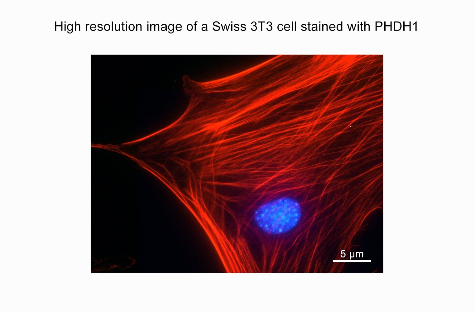
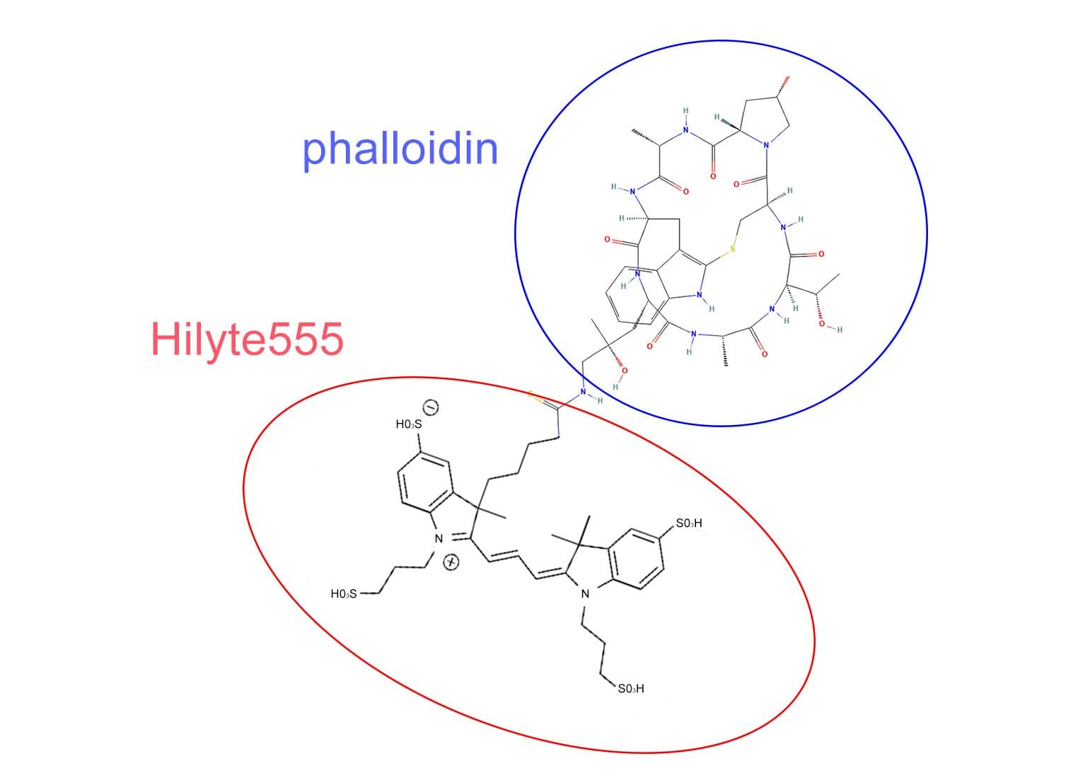
      Phalloidin is a seven amino acid peptide toxin from the mushroom Amanita phalloides, which binds specifically and with high affinity (Kd 20 nM) to the polymerized form of actin (F-actin).  Phalloidin lowers the critical concentration of actin polymerization to less than 1 µg/ml, thereby acting as a polymerization enhancer. Phalloidin has been labeled with a proprietary red fluorescent dye which allows it to be used to stain actin filaments in tissue cultured cells and tissue sections (1, see Figure 1) and cell-free preparations.  Acti-stain™ 555 phalloidin-labeled actin filaments retain many functional characteristics of unlabeled actin including their ability to interact with myosin.  Actin-stain™ 555 phalloidin is supplied as a red solid. 

      Note: Phalloidin is toxic and must be handled with care (LD50 human = 2mg/Kg).


      Example Results and Specifications

      Figure 1.   Actin Stress Fibers stained with Acti-stain™ 555 in a Swiss 3T3 cell.


      PHDH1_img1

      Swiss 3T3 cell with activated Rho, nucleus is stained with Dapi (blue) and F-actin is stained with Acti-stain™ 555 (red F-actin, Cat.# PHDH1).


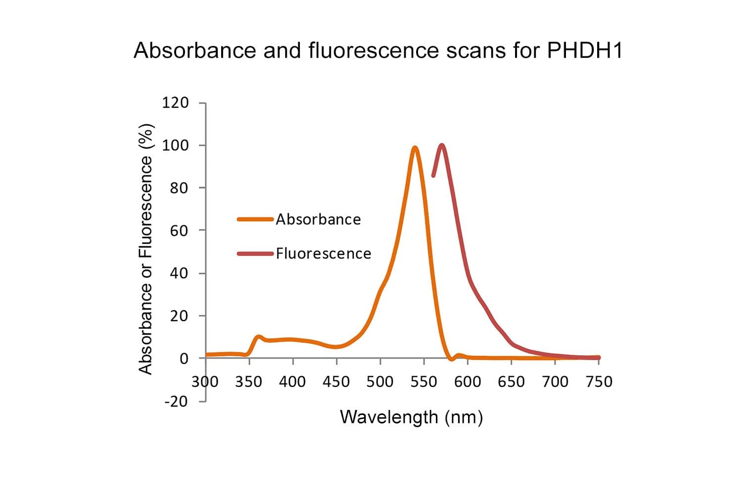
      Figure 2. Emission and excitation scans for Acti-stain™ phalloidins


      PHDH1_scan

      Absorbance and fluorescence scan of Acti-stain™ 555. Labeled phalloidin was diluted into methanol and its absorbance and excitation spectra were scanned between 300-750 and 560-750 nm, respectively. Absorbance peaks at 560 nm and fluorescence at 575 nm.


      Acti-stain phalloidins are the most well characterized phalloidins available. Tabe 1 describes their brightness, photostability, background and affinity constants to F-actin. Compare these performance characteristics to other fluorescent phalloidins and you will see the advantages of using Acti-stain™ for your actin staining requirements.

      Table 1. Biochemical characteristics of fluorescent phalloidins

      ConjugateCat.#

      Wavelengths (Ex/Em)

      Brightness (AFU*)

      Stability to photobleaching(half life in seconds**)

      Background (% of total AFU at 100nM**)

      Affinity (Kd in nanomolar)

      Fluorescein-phalloidin

      na485/535 FITC filter set43262272 +/-12

      Acti-stain™ 488

      PHDG1485/535 FITC filter set83257555 +/-8

      Acti-stain™ 535

      PHDR1535/585 TRITC filter set430271236 +/-7

      Acti-stain™ 555

      PHDH1535/585 TRITC filter set551461663 +/-8

      Acti-stain™ 670

      PHDN1640/680 Cy5 filter set33281850 +/-12

      * = AFU's measured by quantitative cell imaging. ** = Measured in stained Swiss 3T3 cells in the absence of antifade.


      For more information about protocols to fix and stain cells click here.


      References

      1. Wulf, E. et al. (1979). Proc Natl Acad Sci USA. 76(9):4498-4502.

      2. Kron, S.J. et al. (1991).  Meth. Enzmol. 196: 399-416.

      For product Datasheets and MSDSs please click on the PDF links below.   For additional information, click on the FAQs tab above or contact our Technical Support department at tservice@cytoskeleton.com

      AuthorTitleJournalYearArticle Link
      Bhende, Prajakta Praveen et al.Haloarchaeal poly[(3-hydroxybutyrate)- co -(3-hydroxyvalerate)] composite films reinforced with graphene nanoplatelets as a biomaterial for skin tissue engineeringRSC Advances2024
      Vieira, Tânia et al.Electrospun poly(lactic acid) membranes with defined pore size to enhance cell infiltrationHeliyon2024
      Liu, Xiao et al.Erythropoietin regulates osteoclast formation via up-regulating PPARγ expressionMolecular medicine2024
      Fisher, Lucas A.B. et al.Filamin protects myofibrils from contractile damage through changes in its mechanosensory regionPLOS Genetics2024
      Lozano-Vidal, Noelia et al.Aging-regulated PNUTS maintains endothelial barrier function via SEMA3B suppressionCommunications Biology2024
      Tamura, Isao et al.Nuclear actin assembly is an integral part of decidualization in human endometrial stromal cellsCommunications Biology2024
      Wang, Xing et al.RPN1: a pan-cancer biomarker and disulfidptosis regulatorTranslational Cancer Research2024
      Oyama, Shohei et al.Cyclic compressive loading induces a mature meniscal cell phenotype in mesenchymal stem cells with an atelocollagen-based scaffoldFrontiers in Bioengineering and Biotechnology2024
      Zhang, Shiyu et al.PPARγ Antagonists Exhibit Antitumor Effects by Regulating Ferroptosis and DisulfidptosisBiomolecules2024
      Shimabukuro, Masaya et al.Antibacterial and osteogenic thin films on Ti-6Al-4V surface formed by passivation process in copper hydroxide solutionScience and Technology of Advanced Materials2024
      Iwamoto, Yoshiaki et al.Nannostelium ampullaceum gen. et sp. nov., a tiny new member of the protosteloid amoeba of the Cavosteliida (Variosea, Amoebozoa)Protist2024
      Li, Xue et al.Colorectal cancer cells secreting DKK4 transform fibroblasts to promote tumour metastasisOncogene2024
      Clements, Rhiannon et al.Myosin XV is a negative regulator of signaling filopodia during long-range lateral inhibitionDevelopmental Biology2024
      Huang, Zhengqi et al.Faster structured illumination microscopy using complementary encoding-based compressive imagingPhotonics Research2024
      Tang, Jun et al.Disulfidptosis-related signature predicts prognosis and characterizes the immune microenvironment in hepatocellular carcinomaCancer Cell International2024
      Zhang, Qianqian et al.Lithium-Doped Titanium Dioxide-Based Multilayer Hierarchical Structure for Accelerating Nerve-Induced Bone RegenerationACS Applied Materials and Interfaces2024
      Ren, Xu et al.Differences in F pocket impact on HLA I genetic associations with autoimmune diabetesFrontiers in Immunology2024
      Sakagami, Tosuke et al.Structure of putative epidermal sensory receptors in an acoel flatworm, Praesagittifera naikaiensisCell and Tissue Research2024
      Pastorino, Gil A. et al.A partial epithelial-mesenchymal transition signature for highly aggressive colorectal cancer cells that survive under nutrient restrictionThe Journal of Pathology2024
      Kimura, Ken Ichi et al.Activin is a neural inducer of a male-specific muscle in DrosophilaScientific Reports 2024
      Noda, Kazuya et al.Characterization of Rab32- and Rab38-positive lysosome-related organelles in osteoclasts and macrophagesJournal of Biological Chemistry2023
      Shirakawa, Makoto et al.Visualization of root extracellular traps in an ectomycorrhizal woody plant (Pinus densiflora) and their interactions with root-associated bacteriaPlanta2023
      Massaro, Matteo et al.Lipid nanoparticle-mediated mRNA delivery in lung fibrosisEuropean Journal of Pharmaceutical Sciences2023
      Wu, Peizheng et al.Unusual effects of a nanoporous gold substrate on cell adhesion and differentiation because of independent multi-branch signaling of focal adhesionsJournal of Materials Science: Materials in Medicine2023
      Pires, Laura S. et al.PEDOT-Coated PLA Fibers Electrospun from Solutions Incorporating Fe(III)Tosylate in Different Solvents by Vapor-Phase Polymerization for Neural RegenerationPolymers2023
      Turner, Madison et al.Lysosomal swelling and lysis mediate delivery of C3 toxins to their cytoplasmic targetsMolecular Microbiology2023
      Phillips, Andrew T. et al.The formin DAAM1 regulates the deubiquitinase activity of USP10 and integrin homeostasisEuropean Journal of Cell Biology2023
      Boonrod, Kajohn et al.Identification of the Actin-Binding Region and Binding to Host Plant Apple Actin of Immunodominant Transmembrane Protein of ‘Candidatus Phytoplasma mali’International Journal of Molecular Sciences2023
      Nattasit, Praphawi et al.Stiffness-Tunable Hydrogel-Sandwich Culture Modulates the YAP-Mediated Mechanoresponse in Induced-Pluripotent Stem Cell Embryoid Bodies and Augments Cardiomyocyte DifferentiationMacromolecular Bioscience2023
      Sun, Lixiang et al.Dynamic interplay between IL-1 and WNT pathways in regulating dermal adipocyte lineage cells during skin development and wound regenerationCell reports2023
      Wang, Lihui et al.SAYSD1 senses UFMylated ribosome to safeguard co-translational protein translocation at the endoplasmic reticulumCell reports2023
      Shoji, Kenji F et al. The mechanosensitive TRPV2 calcium channel promotes human melanoma invasiveness and metastatic potential EMBO reports2023
      Evangelista, Baggio A. et al.Tandem detergent-extraction and immunoprecipitation of proteinopathy: Scalable enrichment of ALS-associated TDP-43 aggregatesiScience2023
      Ji, Mintao et al.The role of mechano-regulated YAP/TAZ in erectile dysfunctionNature Communications2023
      Li, Dong et al.GxcM-Fbp17/RacC-WASP signaling regulates polarized cortex assembly in migrating cells via Arp2/3Journal of Cell Biology2023
      Cakir, Zeynep et al.Quantitative Proteomic Analysis Reveals apoE4-Dependent Phosphorylation of the Actin-Regulating Protein VASPMolecular and Cellular Proteomics2023
      Awoniyi, Luqman O. et al.B cell receptor-induced protein dynamics and the emerging role of SUMOylation revealed by proximity proteomicsJournal of Cell Science2023
      Hu, Min et al.Defective neurite elongation and branching in Nibp/Trappc9 deficient zebrafish and miceInternational Journal of Biological Sciences2023
      Qin, Yizhi et al.High-Throughput, Low Background, and Wide-Field Microscopy by Flat-Field Photobleaching Imprinting MicroscopyChemical and Biomedical Imaging2023
      Tonami, Kazuo et al.Coordinated linear and rotational movements of endothelial cells compartmentalized by VE-cadherin drive angiogenic sproutingiScience2023
      Karra, N. et al.The effect of membrane properties on cell growth in an ‘Airway barrier on a chip’Organs-on-a-Chip2023
      Baudo, Gherardo et al.Polymer-Functionalized Mitochondrial Transplantation to Fibroblasts Counteracts a Pro-Fibrotic PhenotypeInternational Journal of Molecular Sciences2023
      Bhanja, Anshuman et al.N-WASP-dependent branched actin polymerization attenuates B-cell receptor signaling by increasing the molecular density of receptor clusterseLife2023
      Xia, Qiaoxi et al.A protein complex of LCN2, LOXL2 and MMP9 facilitates tumour metastasis in oesophageal cancerMolecular Oncology2023
      Oh, Mijung et al.High extracellular glucose promotes cell motility by modulating cell deformability and contractility via the cAMP-RhoA-ROCK axis in human breast cancer cellsMolecular Biology of the Cell2023
      Oda, Haruka et al.Actin filaments accumulated in the nucleus remain in the vicinity of condensing chromosomes in the zebrafish early embryoBiology Open2023
      Adhicary, Subhodip et al.Rbpj Deficiency Disrupts Vascular Remodeling via Abnormal Apelin and Cdc42 (Cell Division Cycle 42) Activity in Brain Arteriovenous MalformationStroke2023
      Suzuki, Soichiro et al.ZNF185 prevents stress fiber formation through the inhibition of RhoA in endothelial cellsCommunications Biology2023
      Shoji, Kenji F et al.The mechanosensitive TRPV2 calcium channel promotes human melanoma invasiveness and metastatic potentialEMBO reports2023
      Bioactivity, MPF et al.Bioactivity Enhancement of Plasma-Sprayed Hydroxyapatite Coatings through Non-Contact Corona Electrical ChargingNanomaterials2023
      Haines, A. et al.Chlamydia trachomatis Subverts Alpha-Actinins To Stabilize Its InclusionMicrobiology Spectrum2023
      Sohn, Claira et al.Pathogenic tau decreases nuclear tension in cultured neuronsFrontiers in Aging2023
      O’Shea, T. M. et al.Lesion environments direct transplanted neural progenitors towards a wound repair astroglial phenotype in miceNature Communications2022
      Jain, Ishita et al.Delineating cooperative effects of Notch and biomechanical signals on patterned liver differentiationCommunications Biology2022
      Izumi, Hideki et al.Asymmetric Pericentrosomal CD133 Endosomes Induce the Unequal Autophagic Activity During Cytokinesis in CD133-Positive Human Neuroblastoma CellsStem Cells2022
      Cao, Yu Fei et al.Targeting USP10 induces degradation of oncogenic ANLN in esophageal squamous cell carcinomaCell Death & Differentiation2022
      Music, Amna et al.The SNARE protein Vti1b is recruited to the sites of BCR activation but is redundant for antigen internalisation, processing and presentationFrontiers in Cell and Developmental Biology2022
      Winston, Tackla S. et al.Controlling Mesenchyme Tissue Remodeling via Spatial Arrangement of Mechanical ConstraintsFrontiers in Bioengineering and Biotechnology2022
      Küey, Cansu et al.Recruitment of clathrin to intracellular membranes is sufficient for vesicle formationeLife2022
      Sundby, Lauren J. et al.Nucleotide- and Protein-Dependent Functions of Actg1Molecular Biology of the Cell2022
      Hiratsuka, Ken et al.Organoid-on-a-chip model of human ARPKD reveals mechanosensing pathomechanisms for drug discoveryScience Advances2022
      Du, Qiqiao et al.FASN promotes lymph node metastasis in cervical cancer via cholesterol reprogramming and lymphangiogenesisCell Death & Disease2022
      Gupta, Dhanak et al.Traditional Multiwell Plates and Petri Dishes Limit the Evaluation of the Effects of Ultrasound on Cells In VitroUltrasound in Medicine & Biology2022
      Malin, Jacob et al.Sidekick dynamically rebalances contractile and protrusive forces to control tissue morphogenesisJournal of Cell Biology2022
      Zheng, Lijun et al.Ionizing radiation-induced long noncoding RNA CRYBG3 regulates YAP/TAZ through mechanotransductionCell Death & Disease 2022
      Wang, Ling et al.Novel oral edaravone attenuates diastolic dysfunction of diabetic cardiomyopathy by activating the Nrf2 signaling pathwayEuropean Journal of Pharmacology2022
      Huang, Qichao et al.TFAM loss induces nuclear actin assembly upon mDia2 malonylation to promote liver cancer metastasisThe EMBO Journal2022
      Nanamatsu, Azuma et al.Vas******** Induces Urinary Uromodulin Secretion by Activating PKA (Protein Kinase A)Hypertension2021
      Kimura, Ken ichi et al.Crucial role of framework with cytoskeletal actin filaments for shaping microstructure of footpad setae in the ladybird beetle, Harmonia axyridisArthropod Structure and Development2021
      Shindo, Taiki et al.Visualization of endogenous nuclear F-actin in mouse embryos reveals abnormal actin assembly after somatic cell nuclear transferJournal of biochemistry2021
      Gharanei, Seley et al.Vascular Adhesion Protein-1 Determines the Cellular Properties of Endometrial PericytesFrontiers in Cell and Developmental Biology2021
      Kanamoto, Takashi et al.Integrin α2β1 plays an important role in the interaction between human articular cartilage-derived chondrocytes and atelocollagen gelScientific Reports2021
      Chen, Lena et al.Ankyrin-B p.S646F undergoes increased proteasome degradation and reduces cell viability in the H9c2 rat ventricular cardiomyoblast cell lineBiochem Cell Biol2020
      Chemla, Yoav et al.Carbon nanostructures as a scaffold for human embryonic stem cell differentiation toward photoreceptor precursorsNanoscale2020
      Liu, Yi et al.Effect of F-actin and Microtubules on Cellular Mechanical Behavior Studied Using Atomic Force Microscope and an Image Recognition-Based Cytoskeleton Quantification ApproachInternational Journal of Molecular Sciences2020
      Hamdan, Hamdan et al.Mapping axon initial segment structure and function by multiplexed proximity biotinylationNature Communications2020
      Ding, Dalian et al.Some Ototoxic Drugs Destroy Cochlear Support Cells Before Damaging Sensory Hair CellsNeurotoxicity Research2020
      Pęziński, Marcin et al.An improved method for culturing myotubes on laminins for the robust clustering of postsynaptic machineryScientific Reports2020
      Hall, Everett G. et al.SPECC1L regulates palate development downstream of IRF6Human Molecular Genetics2020
      Ding, Dalian et al.Antioxidative stress-induced damage in cochlear explantsJournal of Otology2020
      Fu, Yi et al.Elevation of JAML Promotes Diabetic Kidney Disease by Modulating Podocyte Lipid MetabolismCell Metabolism2020
      Bokelmann, Marcel et al.Utility of primary cells to examine npc1 receptor expression in mops condylurus, a potential ebola virus reservoirPLoS Neglected Tropical Diseases2020
      Campisi, Marco et al.Tumor-Derived cGAMP Regulates Activation of the VasculatureFrontiers in Immunology2020
      Kimura, Ken ichi et al.Framework with cytoskeletal actin filaments forming insect footpad hairs inspires biomimetic adhesive device designCommunications Biology2020
      Ueta, Miho et al.Effects of TGF-β1 on the migration and morphology of RAW264.7 cells in vitroMolecular Medicine Reports2019
      Ye, Haibo et al.Bilirubin-induced neurotoxic and ototoxic effects in rat cochlear and vestibular organotypic culturesNeuroToxicology2019
      Fultz, Robert et al.Phagocytosis by macrophages depends on histamine H2 receptor signaling and scavenger receptor 1MicrobiologyOpen2019
      Mandigo, Torrey R. et al.Drosophila emerins control LINC complex localization and transcription to regulate myonuclear positionJournal of Cell Science2019
      Ferguson, Sylvia H. et al.Interferon-λ3 Promotes Epithelial Defense and Barrier Function Against Cryptosporidium parvum InfectionCmgh2019
      Siddiqui, Nida et al.PTPN21 and Hook3 relieve KIF1C autoinhibition and activate intracellular transportNature Communications2019
      Vohnoutka, Rishel B. et al.The focal adhesion scaffold protein Hic-5 regulates vimentin organization in fibroblastsMolecular Biology of the Cell2019
      Del Signore, Steven J. et al.The WAVE Regulatory Complex and Branched F-Actin Counterbalance Contractile Force to Control Cell Shape and Packing in the Drosophila EyeDevelopmental Cell2018
      Bardai, Farah H. et al.A conserved cytoskeletal signaling cascade mediates neurotoxicity of FTDP-17 tau mutations in vivoJournal of Neuroscience2018
      Wang, Liqun et al.Tissue and cellular rigidity and mechanosensitive signaling activation in Alexander diseaseNature Communications2018
      Nakamura, Koji et al.Exosomes promote ovarian cancer cell invasion through transfer of CD44 to peritoneal mesothelial cellsMolecular Cancer Research2017
      Della Torre, Camilla et al.Adsorption of B(α)P on carbon nanopowder affects accumulation and toxicity in zebrafish (Danio rerio) embryosEnvironmental Science: Nano2017
      Collins, Mary Ann et al.Emery-Dreifuss muscular dystrophy-linked genes and centronuclear myopathy-linked genes regulate myonuclear movement by distinct mechanismsMolecular Biology of the Cell2017
      Rao, Yanhua et al.A Mec17-Myosin II effector axis coordinates microtubule acetylation and actin dynamics to control primary cilium biogenesisPLoS ONE2014
      Burks, J. et al.ISGylation governs the oncogenic function of Ki-Ras in breast cancerOncogene 2014 33:62013
      Andersen, Thomas E. et al.Escherichia coli uropathogenesis In vitro: Invasion, cellular escape, and secondary infection analyzed in a human bladder cell infection modelInfection and Immunity2012

      Question 1:  Can I use fluorescently-labeled phalloidin to stain F-actin in living cells?

      Answer 1:  Unfortunately, no, phalloidin cannot be used to stain F-actin in living cells.  Phalloidins are used to stain F-actin in fixed cells.  Fluorescent phalloidins only bind to the native quaternary structure of F-actin which provides a low background.  The correct fixation condition for phalloidin binding is 3.7% (v/v) paraformaldehyde in PBS for 10 min because it retains the quaternary protein structure which is necessary for high affinity binding of phalloidin.  Methanol fixation destroys the native conformation and hence is not suitable for F-actin staining with phalloidin.  To monitor actin dynamics in living cells, micro-injection of rhodamine-labeled actin (Cat. # APHR or AR05) is recommended.  Please see those datasheets for more information.


      Question 2:  Which of the fluorescently-labeled phalloidin is the most stable/brightest?

      Answer 2:  The brightest and most stable of the Acti-stains is Acti-stain 488 (green fluorescence; Cat. # PHDG1).  Please see the table below for additional information on all of our Acti-stains. 

      Conjugate

      Cat. #

      Wavelengths (Ex/EM)

      Brightness (AFU)

      Stability to photobleaching* (1/2 life in sec)

      Background (% of total AFU at 100 nM)

      Acti-stain™ 488

      PHDG1

      485/535

      832

      57

      5

      Acti-stain™ 535

      PHDR1

      535/585

      430

      27

      12

      Acti-stain™ 555

      PHDH1

      535/585

      551

      46

      16

      Acti-stain™ 670

      PHDN1

      640/680

      332

      18

      18

      * = measured in the absence of antifade.


      If you have any questions concerning this product, please contact our Technical Service department at tservice@cytoskeleton.com.