SiR-DNA Kit

SiR-DNA Kit
$0.00
SKU
CY-SC007

SiR-DNA is a far-red, fluorogenic, cell permeable, low background and highly specific probe for DNA. SiR-DNA is based on the DNA minor groove binder bisbenzimide, it allows the labelling of DNA in live cells with high specificity and low background. SiR-DNA is fluorogenic, cell permeable and highly specific for DNA. Sir-DNA stains the nuclei of live cells without the need for genetic manipulation or overexpression. Its emission in the far red minimizes phototoxicity and sample autofluorescence. This reagent has multiple advantages (far-red, fluorogenic, cell permeable, low background and highly specific probe for DNA) over existing probes such Syto61, DRAQ5 or Vybrant DyeCycle Ruby dyes.

SiR-DNA is compatible with GFP and/or m-cherry fluorescent proteins. It can be imaged with standard Cy5 filtersets. SiR-actin can be used for widefield, confocal, SIM or STED imaging in living cells and tissue. Probe quantity allows 50 – 300 staining experiments.*

For comparison information with other nuclear stains click here.
 

Optical properties

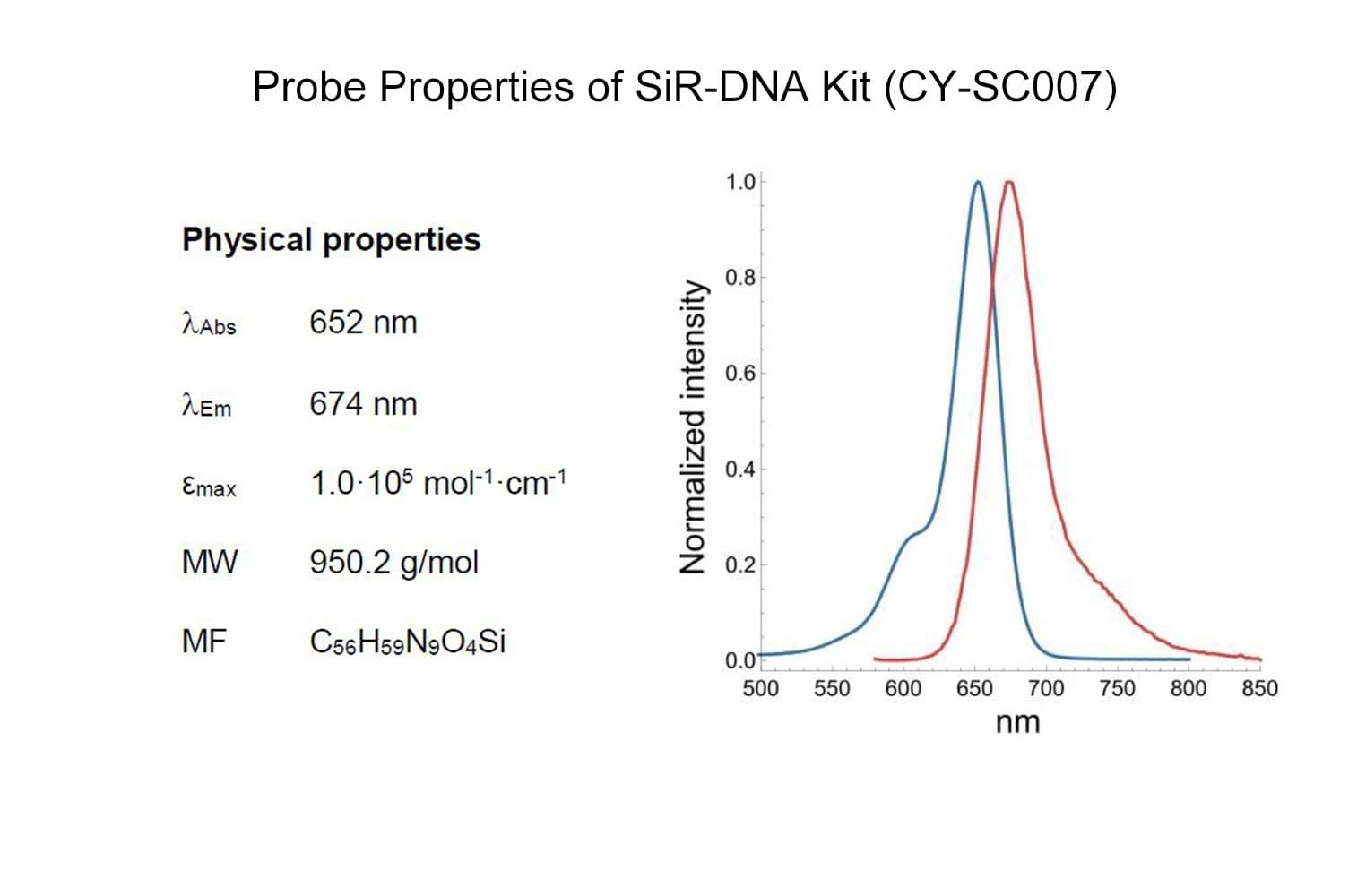
λabs   652 nm

λEm   674 nm
εmax 1.0·105 mol-1·cm-1


*Based on the following conditions: 0.3 – 1 ml staining solution / staining experiments with 0.5 – 1 µM probe concentration. The number of staining experiments can be further increased by reducing volume or probe concentration.


Cytoskeleton, Inc. is the exclusive provider of Spirochrome, Ltd. products in North America.

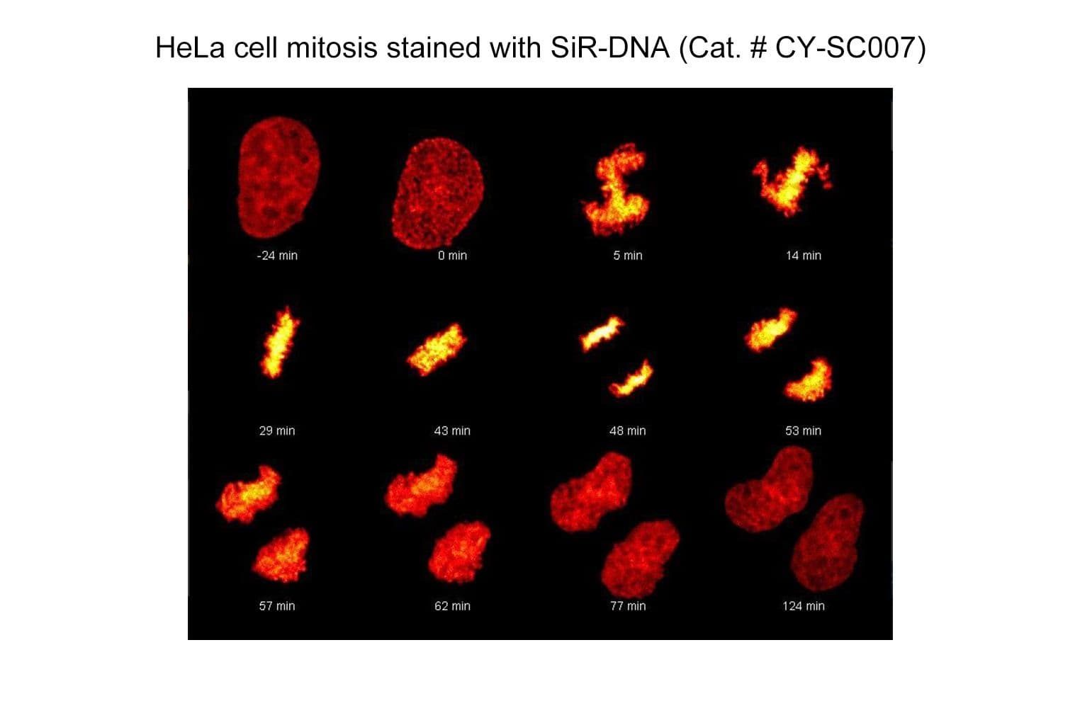
Live cell time lapse confocal imaging of a HeLa cell stained with SiR-DNA, note the very low background.

For product Datasheets and MSDSs please click on the PDF links below.

For comparison information with other nuclear stains click here.

AuthorTitleJournalYearArticle Link
Wang, Chao et al.Protocol for the detection of mechanosensitive response of proteins in cultured cells under compressive stressSTAR Protocols2024
Zhang, Wenya et al.Protocol for culturing and functionally manipulating planarian neoblasts using SiR-DNA-based flow cytometrySTAR Protocols2024
Daugird, Timothy A. et al.Correlative single molecule lattice light sheet imaging reveals the dynamic relationship between nucleosomes and the local chromatin environmentNature Communications 2024 15:12024
Richter, Manuela et al.Kinetochore-fiber lengths are maintained locally but coordinated globally by poles in the mammalian spindleeLife2023
Zeeshan, Mohammad et al.Plasmodium ARK2 and EB1 drive unconventional spindle dynamics, during chromosome segregation in sexual transmission stagesNature Communications 2023 14:12023
Sterling, Noelle A. et al.An entosis-like process induces mitotic disruption in Pals1 microcephaly pathogenesisNature Communications 2023 14:12023
Slangen, P L G et al.Protocol for live-cell imaging during Tumor Treating Fields treatment with Inovitro LiveSTAR protocols2022
Veling, Mike T. et al.Natural and Designed Proteins Inspired by Extremotolerant Organisms Can Form Condensates and Attenuate Apoptosis in Human CellsACS Synthetic Biology2022
Samejima, Itaru et al.Mapping the invisible chromatin transactions of prophase chromosome remodelingMolecular Cell2022
Rashid, Fatema Zahra M. et al.HI-NESS: a family of genetically encoded DNA labels based on a bacterial nucleoid-associated proteinNucleic acids research2022
Houthaeve, Gaëlle et al.Transient nuclear lamin A/C accretion aids in recovery from vapor nanobubble-induced permeabilisation of the plasma membraneCellular and Molecular Life Sciences2022
Safieddine, Adham et al.A choreography of centrosomal mRNAs reveals a conserved localization mechanism involving active polysome transportNature Communications 2021 12:12021
Haroon, Mohammad et al.Myofiber stretch induces tensile and shear deformation of muscle stem cells in their native nicheBiophysical Journal2021
Tsuchiya, Kenta et al.Ran-GTP Is Non-essential to Activate NuMA for Mitotic Spindle-Pole Focusing but Dynamically Polarizes HURP Near ChromosomesCurrent Biology2021
Menegakis, Apostolos et al.Resistance of Hypoxic Cells to Ionizing Radiation Is Mediated in Part via Hypoxia-Induced QuiescenceCells2021
Noa, Amra et al.The hierarchical packing of euchromatin domains can be described as multiplicative cascadesPLOS Computational Biology2021
Zhao, Bing et al.Optogenetic Control of Myocardin-Related Transcription Factor A Subcellular Localization and Transcriptional Activity Steers Membrane Blebbing and Invasive Cancer Cell MotilityAdvanced Biology2021
Safaralizade, Mahira et al.Measuring nuclear calcium and actin assembly in living cellsJournal of biochemistry2021
Grosser, Steffen et al.Cell and Nucleus Shape as an Indicator of Tissue Fluidity in CarcinomaPhysical Review X2021
Geraghty, Zoe et al.The association of Plk1 with the astrin-kinastrin complex promotes formation and maintenance of a metaphase plateJournal of Cell Science2021
Buchmann, B. et al.Mechanical plasticity of collagen directs branch elongation in human mammary gland organoidsNature Communications 2021 12:12021
Scheffler, Kathleen et al.Two mechanisms drive pronuclear migration in mouse zygotesNature Communications 2021 12:12021
Geoghegan, Niall D. et al.4D analysis of malaria parasite invasion offers insights into erythrocyte membrane remodeling and parasitophorous vacuole formationNature Communications 2021 12:12021
Scott, Stacey J. et al.Evidence that polyploidy in esophageal adenocarcinoma originates from mitotic slippage caused by defective chromosome attachmentsCell Death & Differentiation 2021 28:72021
Kurzbauer, Marie Therese et al.ATM controls meiotic DNA double-strand break formation and recombination and affects synaptonemal complex organization in plantsThe Plant Cell2021
Singh, Divya et al.Destabilization of Long Astral Microtubules via Cdk1-Dependent Removal of GTSE1 from Their Plus Ends Facilitates Prometaphase Spindle OrientationCurrent Biology2021
Jagrić, Mihaela et al.Optogenetic control of prc1 reveals its role in chromosome alignment on the spindle by overlap length-dependent forceseLife2021
Bufe, Anja et al.Wnt signaling recruits KIF2A to the spindle to ensure chromosome congression and alignment during mitosisProceedings of the National Academy of Sciences of the United States of America2021
Burigotto, Matteo et al.Centriolar distal appendages activate the centrosome‐PIDDosome‐p53 signalling axis via ANKRD26The EMBO Journal2021
Chen, Jiji et al.Three-dimensional residual channel attention networks denoise and sharpen fluorescence microscopy image volumesNature Methods2021
de Man, S. M.A. et al.Quantitative live-cell imaging and computational modelling shed new light on endogenous wnt/ctnnb1 signaling dynamicseLife2021
Gryaznova, Yulia et al.Kinetochore individualization in meiosis I is required for centromeric cohesin removal in meiosis IIThe EMBO Journal2021
Papini, Diana et al.The Aurora B gradient sustains kinetochore stability in anaphaseCell Reports2021
Harkes, Rolf et al.Dynamic FRET-FLIM based screening of signal transduction pathwaysScientific Reports2021
Wu, Xi et al.A free-form patterning method enabling endothelialization under dynamic flowBiomaterials2021
Hilbert, Lennart et al.Transcription organizes euchromatin via microphase separationNature Communications2021
Ito, Kei K. et al.Cep57 and Cep57L1 maintain centriole engagement in interphase to ensure centriole duplication cycleJournal of Cell Biology2021
Cordero-Espinoza, Lucía et al.Dynamic cell contacts between periportal mesenchyme and ductal epithelium act as a rheostat for liver cell proliferationCell Stem Cell2021
Vukušić, Kruno et al.Microtubule-sliding modules based on kinesins EG5 and PRC1-dependent KIF4A drive human spindle elongationDevelopmental Cell2021
Warmt, Enrico et al.Differences in cortical contractile properties between healthy epithelial and cancerous mesenchymal breast cellsNew Journal of Physics2021
Nowak-Imialek, Monika et al.In Vitro and in Vivo Interspecies Chimera Assay Using Early Pig EmbryosCellular Reprogramming2020
Chinen, Takumi et al. Nu MA assemblies organize microtubule asters to establish spindle bipolarity in acentrosomal human cells The EMBO Journal2020
Benedict, Bente et al.WAPL-Dependent Repair of Damaged DNA Replication Forks Underlies Oncogene-Induced Loss of Sister Chromatid CohesionDevelopmental Cell2020
Roscioli, Emanuele et al.Ensemble-Level Organization of Human Kinetochores and Evidence for Distinct Tension and Attachment SensorsCell Reports2020
Hégarat, Nadia et al.Cyclin A triggers Mitosis either via the Greatwall kinase pathway or Cyclin BThe EMBO Journal2020
Pribyl, Miroslav et al.Aberrantly elevated suprabasin in the bone marrow as a candidate biomarker of advanced disease state in myelodysplastic syndromesMolecular Oncology2020
Andrieu, Cyril et al.MMP14 is required for delamination of chick neural crest cells independently of its catalytic activityDevelopment (Cambridge)2020
Djeghloul, Dounia et al.Identifying proteins bound to native mitotic ESC chromosomes reveals chromatin repressors are important for compactionNature Communications2020
Watts, Lotte P. et al.The rif1-long splice variant promotes g1 phase 53bp1 nuclear bodies to protect against replication stresseLife2020
van der Lelij, Petra et al.STAG1 vulnerabilities for exploiting cohesin synthetic lethality in STAG2-deficient cancersLife Science Alliance2020
Valenzuela, José Ignacio et al.Localized Intercellular Transfer of Ephrin-As by Trans-endocytosis Enables Long-Term SignalingDevelopmental Cell2020
Plessner, Matthias et al.Centrosomal Actin Assembly Is Required for Proper Mitotic Spindle Formation and Chromosome CongressioniScience2019
Dudka, Damian et al.Spindle-Length-Dependent HURP Localization Allows Centrosomes to Control Kinetochore-Fiber Plus-End DynamicsCurrent Biology2019
van Husen, Lea S. et al.Dual Bioorthogonal Labeling of the Amyloid-β Protein Precursor Facilitates Simultaneous Visualization of the Protein and Its Cleavage ProductsJournal of Alzheimer's disease : JAD2019
Alex, Amal et al.Electroporated recombinant proteins as tools for in vivo functional complementation, imaging and chemical biologyeLife2019
Watanabe, Koki et al.The Cep57-pericentrin module organizes PCM expansion and centriole engagementNature Communications2019
Elbatsh, Ahmed M.O. et al.Distinct Roles for Condensin's Two ATPase Sites in Chromosome CondensationMolecular Cell2019
Imrichova, Terezie et al.Dynamic PML protein nucleolar associations with persistent DNA damage lesions in response to nucleolar stress and senescence-inducing stimuliAging2019
Holzmann, Johann et al.Absolute quantification of Cohesin, CTCF and their regulators in human cellseLife2019
Liccardi, Gianmaria et al.RIPK1 and Caspase-8 Ensure Chromosome Stability Independently of Their Role in Cell Death and InflammationMolecular Cell2019
Janssen, Louise M.E. et al.Loss of Kif18A Results in Spindle Assembly Checkpoint Activation at Microtubule-Attached KinetochoresCurrent Biology2018
Avogaro, Laura et al.Live-cell imaging reveals the dynamics and function of single-telomere TERRA molecules in cancer cellsRNA Biology2018
Burgess, Selena G et al.Mitotic spindle association of TACC3 requires Aurora‐A‐dependent stabilization of a cryptic α‐helixThe EMBO Journal2018
Nguyen, Marie et al.Dissecting Effects of Anti-cancer Drugs and Cancer-Associated Fibroblasts by On-Chip Reconstitution of Immunocompetent Tumor MicroenvironmentsCell Reports2018
Politi, Antonio Z. et al.Quantitative mapping of fluorescently tagged cellular proteins using FCS-calibrated four-dimensional imagingNature Protocols2018
Yuan, Ruixue et al.Chk1 and 14‐3‐3 proteins inhibit atypical E2Fs to prevent a permanent cell cycle arrestThe EMBO Journal2018
Walther, Nike et al.A quantitative map of human Condensins provides new insights into mitotic chromosome architectureJournal of Cell Biology2018
Koch, Birgit et al.Generation and validation of homozygous fluorescent knock-in cells using CRISPR-Cas9 genome editingNature Protocols2018
Neubert, Elsa et al.Chromatin swelling drives neutrophil extracellular trap releaseNature Communications2018
Okumura, Masako et al.Dynein–dynactin–NuMA clusters generate cortical spindle-pulling forces as a multiarm ensembleeLife2018
Kubitschke, H. et al.Actin and microtubule networks contribute differently to cell response for small and large strainsNew Journal of Physics2017

Q1. What are the filter sets for the SiR probes?

A1. The SiR actin and tubulin probes are visualized with standard Cy5 filters.  Optimal excitation is 650 nm and emission is 670 nm.  We recommend filters with an excitation of 630 + 20 nm and an emission of 680 + 20 nm (Fig. 1).  

faq-fig3

Figure 1. Excitation (blue) and emission (red) spectra for SiR probes.

Q2. Why do the SiR probes have a low background compared to other fluorophores?

A4. SiR probes are excited by and emit light in the near infrared/far red spectral range, thus avoiding the use of shorter wavelengths such as blue and green light that typically autofluoresce, causing higher background signals.  SiR-coupled probes possess two physical states: 1. a non-fluorescent, closed off-state (spirolactone)  and 2. an open, highly fluorescent on-state (zwitterion).  The binding of the probe to its ligand target favors the highly fluorescent open state while the free unbound probe exists in the closed, non-fluorescent state (Fig. 2).  The fluorescence amplification is 100-fold from the unbound to bound state. This results in a highly sensitive biosensor in which the majority of fluorescence occurs only in the bound state (see Refs. 3 and 4). 

faq-fig4

Figure 2. SiR derivatives exist in equilibrium between the fluorescent zwitterionic (open) form (left structure) and the non-fluorescent spiro (closed) form (right structure).

For comparison information with other nuclear stains click here.

Q3: Are the SiR probes stable at room temperature?

A3: Yes, the probes are stable at room temperature for a few days.  However, it strongly depends on the probe and the solvent.  Thus, it is recommended to store all of the probes or solutions at –20°C.


Q4: Are SiR-DNA, SiR-actin and SiR-tubulin toxic to cells?

A4: Yes, above a certain threshold both probes show some effect on cell proliferation and altered actin or microtubule dynamics.  However, the probes are orders of magnitude less toxic than their parent drug.  In HeLa cells, neither actin nor microtubule dynamics were altered at concentrations below 100 nM.  At this concentration, SiR probes efficiently label microtubules and F-actin, allowing for the capture of high signal to noise images.


Q5: Do the probes work on fixed cells?

A5: SiR-DNA labels all types of fixed cells. SiR-actin probes can be used with PFA-fixed cells.  SiR-actin labels F-actin in PFA-fixed cells as efficiently as phalloidin derivatives.  SiR-tubulin labels microtubules only in ethyleneglycol-bis-succinimidyl-succinate (EGS)-fixed cells.  However, a selective labeling of centrosomal microtubules of PFA-fixed cells was observed.  SiR-actin and SiR-tubulin are not suitable for methanol-fixed cells.


Q6: Is it possible to image SiR-probes by STORM?

A6: No—under the very high light intensities typically used in STORM imaging, a phototoxic effect is observed on live cells.


Q7: Which organisms and tissues are stained by SiR-probes?

A7: This list describes only cell lines, tissues, or organisms that have been reported to work.  Omission of a cell line, tissue, or organism does not mean that the SiR-probes will not work with the specific cells, tissues, or organisms.


Homo sapiens: U2OS, fibroblasts, HeLa, HUVEC, MCF-10A, HCT-116, A549, erythrocytes

Mus musculus: C2C12, IA32, skeletal muscle, primary cardiomyocyte, primary oocyte

Rattus norvegicus: primary hippocampal neurons, primary cortical neurons, NRK

Cercopithecus aethiops: COS-7

Mesocricetus auratus: BHK

Drosophila melanogaster: Notum epithelium, S2

Didelphis marsupialis: OK cells


Q8. Do SiR-probes work in 3D cell cultures?

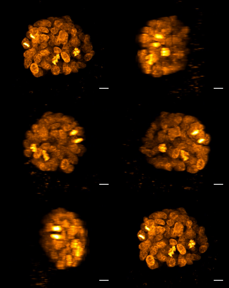
A8: Yes, the probes are able to stain cells in a 3D growth environment.


Q9: What are the correction factors CF260 and CF280 for the SiR fluorophore?

A11: CF260 = 0.116 and CF280  = 0.147

Q10. What is STED microscopy and how does it work?

A10. STED microscopy stands for Stimulated Emission Depletion microscopy.  It is one type of super resolution microscopy which allows the capture of images with a higher resolution than conventional light microscopy which is constrained by diffraction of light.  STED uses 2 laser pulses, one is the excitation pulse which excites the fluorophore, causing it to fluoresce.  The second pulse, referred to as the STED pulse, de-excites the fluorophore via stimulated emission in an area surrounding a central focal spot that is not de-excited and thus continues to fluoresce.  This is accomplished by focusing the STED pulse into a ring shape, a so-called donut, where the center focal spot is devoid of the STED laser pulse, conferring high resolution to the fluorescent area (Fig. 3; see Ref. 1 for more details on STED microscopy).


fig1-faq

Figure 3. STED microscopic image of microtubules labeled with SiR-tubulin in human primary dermal fibroblasts.

Q11. Why is the SiR actin (or tubulin) probe good for STED microscopy?

A11. STED microscopy offers the ability to study cellular details on a nanometermolar scale in vivo.  To take advantage of this super resolution microscopy, one must be able to select with high specificity the area to be examined using fluorescent probes.  In addition, the fluorescent probes must be bright, photostable, exhibit no or little phototoxicity, be excited and emit in the far red spectrum.  In addition, if the probe is to be used for live cell imaging (thus avoiding fixation artifacts that occur when cells are fixed), high cell permeability is necessary.  The SiR-DNA, -actin and -tubulin probes fulfill all of these requirements.  In short, the combination of STED and SiR probes allows for unparalleled fluorescent visualization of subcellular actin and tubulin/microtubule structures and their physical characterization in living cells, (see Fig. 4 and Ref. 2). 

Neuron_actin_1
Neuron_actin_closup2_1

Figure 4. STED images of cultured rat hippocampal neurons stained with SiR-actin. Bottom image is a close-up view of part of the top image to clearly visualize actin rings (stripes) with 180 nm periodicity. Courtesy Of Elisa D'Este, MPI Biophysical Chemistry, Göttingen.

For comparison information with other nuclear stains click here.

References

1. Hell S.W. and Wichmann J. 1994. Breaking the diffraction resolution limit by stimulated emission: stimulated-emission-depletion fluorescence microscopy. Opt. Lett. 19, 780-782.

2. D’Este E. et al. 2015. STED nanoscopy reveals the ubiquity of subcortical cytoskeleton periodicity in living neurons. Cell Rep. 10, 1246-1251.

3. Lukinavicius G. et al. 2013. A near-infrared fluorophore for live-cell super-resolution microscopy of cellular proteins. Nat. Chem. 5, 132-139.

4. Lukinavicius G. et al. 2014. Fluorogenic probes for live-cell imaging of the cytoskeleton. Nature Methods. 11, 731-733.