Actin Binding Protein Spin-Down Assay Biochem Kit: human platelet actin

Actin Binding Protein Spin-Down Assay Biochem Kit: human platelet actin
$0.00

Product Uses Include

  • To determine whether a protein binds to filaments or monomers of actin.
  • To determine whether a protein bundles F-actin
  • To test various conditions (e.g. pH optima) or requirements (e.g. divalent cation requirement) for binding to actin
bk013

Introduction
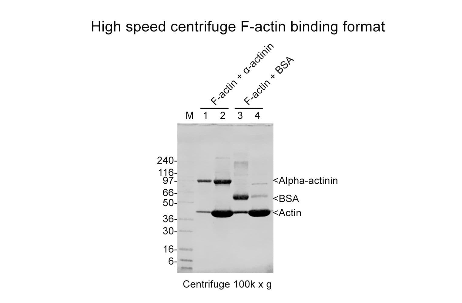
The actin binding protein spin-down assay kit provides G- or F-actin plus positive (α-actinin ) and negative (Bovine Serum Albumin, BSA) binding control proteins. Actin binding occurs when there is an affinity for any site of actin. F-actin binding can be measured by using a spin down method. In this way centrifugation is used to separate F-actin from G-actin by differential sedimentation. F-actin binding proteins will co-sediment with actin filaments and form a pellet at the bottom of the centrifugation tube. Proteins with F-actin bundling activity can be detected since they will cause the F-actin to sediment at lower centrifugal forces than normal (14,000 x g vs 150,000 x g). F-actin severing proteins, G-actin binding proteins or non-actin binding proteins will stay in the supernatant. Severing proteins will be expected if more G-actin remains in the supernatant than in the negative control sample, and this activity should be tested further by measuring F-actin length distributions before and after adding the test protein. G-actin binding proteins can be measured by adding the test protein to G-actin and inducing polymerization, if the test protein sequesters G-actin then during the spin-down assay more actin will be left in the supernatant compared with the control.

Actin can exist in two forms: Globular subunit (G-actin) and Filamentous polymer (F-actin) (See the About Actin page for more information). Both forms of actin interact with a plethora of proteins in the cell. To date there are over 50 distinct classes of Actin-Binding Proteins (ABPs), and the inventory is still far from complete. Actin Binding Proteins allow the actin cytoskeleton to respond rapidly to cellular and extracellular signals and are integral to cytoskeletal involvement in many cellular processes. These include cell shape and motility, muscle contraction, intracellular trafficking, cell pathogenesis and signal transduction.

This kit contains non-muscle actin (Cat. # APHL99). The same kit is also available with skeletal muscle actin (Cat. # AKL99), see Cat. # BK001. The skeletal muscle actin spin-down kit may be more appropriate to use to study actin binding proteins from muscle tissues.

Kit contents
The kit contains sufficient materials for 30-100 assays depending on assay volume. The following reagents are included:

  1. 8 x 250 µg Non-muscle actin (Cat. # APHL99).
  2. α-Actinin, positive control (Cat. # AT01).
  3. BSA, negative control
  4. General Actin Buffer (Cat. # BSA01).
  5. Actin Polymerization Buffer (Cat.# BSA02)
  6. F-actin Cushion Buffer.
  7. ATP, 100 mM (Cat. # BSA04)
  8. EGTA, 0.5 M
  9. MgCl2, 100 mM
  10. Tris-HCl pH 6.5, 1.0 M
  11. Tris-HCl pH 7.5, 100 mM
  12. Manual with detailed protocols and extensive troubleshooting guide

Equipment needed

  1. Centrifugation set-up capable of 150,000 x g at 4°C and 24°C, 50 -200 µl volume capacity.
  2. SDS-PAGE system.
  3. Detection system for protein of interest (coomassie is good for purified proteins, Western blot or silver stain for less pure or low abundance test proteins).
  4. Gel scanner for densitometric determinations.

For product Datasheets and MSDSs please click on the PDF links below.   For additional information, click on the FAQs tab above or contact our Technical Support department at tservice@cytoskeleton.com

AuthorTitleJournalYearArticle Link
Joseph, Jesvin et al.Nanoscale chemical characterization of secondary protein structure of F-Actin using mid-infrared photoinduced force microscopy (PiF-IR)Spectrochimica Acta Part A: Molecular and Biomolecular Spectroscopy2024
Chanez-Paredes, Sandra D. et al.Mechanisms underlying distinct subcellular localization and regulation of epithelial long myosin light-chain kinase splice variantsJournal of Biological Chemistry2024
Szpak, Dorota et al.Kindlin-3 deficiency leads to impaired erythropoiesis and erythrocyte cytoskeletonBlood Advances2023
DeGiosio, R. A. et al.Differential regulation of MAP2 by phosphorylation events in proline-rich versus C-terminal domainsThe FASEB Journal2023
Duan, Xudong et al.Essential role of the endocytic site-associated protein Ecm25 in stress-induced cell elongationCell Reports2021
Yan, Yanling et al.RTKN-1/rhotekin shields endosome-associated F-actin from disassembly to ensure endocytic recyclingJournal of Cell Biology2021
Lu, Yi Ju et al.Arabidopsis calcium-dependent protein kinase 3 regulates actin cytoskeleton organization and immunityNature Communications2020
Park, Jin Suk et al.Mechanical regulation of glycolysis via cytoskeleton architectureNature2020
Gabel, Marion et al.Phosphorylation cycling of Annexin A2 Tyr23 is critical for calcium-regulated exocytosis in neuroendocrine cellsBiochimica et Biophysica Acta - Molecular Cell Research2019
Afreen, Sana et al.Altered Cytoskeletal Composition and Delayed Neurite Elongation in tau45–230-Expressing Hippocampal NeuronsNeuroscience2019
Krueger, Daniel et al.Cross-linker–mediated regulation of actin network organization controls tissue morphogenesisJournal of Cell Biology2019
Mao, Bai Ping et al.CaMSAP2 is a microtubule minus-end targeting protein that regulates BTB dynamics through cytoskeletal organizationEndocrinology2019
Zhang, Min et al.Axonogenesis Is Coordinated by Neuron-Specific Alternative Splicing Programming and Splicing Regulator PTBP2Neuron2019
Wen, Qing et al.Actin nucleator Spire 1 is a regulator of ectoplasmic specialization in the testisCell Death and Disease2018
Bardai, Farah H. et al.A conserved cytoskeletal signaling cascade mediates neurotoxicity of FTDP-17 tau mutations in vivoJournal of Neuroscience2018
Kschonsak, Yvonne T. et al.Activated ezrin controls MISP levels to ensure correct NuMA polarization and spindle orientationJournal of Cell Science2018
Leelarasamee, Natthanon et al.The root-knot nematode effector MiPFN3 disrupts plant actin filaments and promotes parasitismPLoS Pathogens2018
Gong, Ting et al. PTRN ‐1/ CAMSAP promotes CYK ‐1/formin‐dependent actin polymerization during endocytic recycling The EMBO Journal2018
Shioda, Norifumi et al.Targeting G-quadruplex DNA as cognitive function therapy for ATR-X syndrome articleNature Medicine2018
Loukil, Abdelhalim et al.Foci of cyclin A2 interact with actin and RhoA in mitosisScientific Reports2016
Feng, Di et al.Functional validation of an alpha-actinin-4 mutation as a potential cause of an aggressive presentation of adolescent focal segmental glomerulosclerosis: Implications for genetic testingPLoS ONE2016
Gao, Ying et al.Polarity protein Crumbs homolog-3 (CRB3) regulates ectoplasmic specialization dynamics through its action on F-actin organization in Sertoli cellsScientific Reports2016
Wang, Peixiang et al.RAB-10 Promotes EHBP-1 Bridging of Filamentous Actin and Tubular Recycling EndosomesPLoS Genetics2016
Tie, S. R. et al.Regulation of sarcoma cell migration, invasion and invadopodia formation by AFAP1L1 through a phosphotyrosine-dependent pathwayOncogene2016
Li, Nan et al.Overexpression of plastin 3 in Sertoli cells disrupts actin microfilament bundle homeostasis and perturbs the tight junction barrierSpermatogenesis2016
Gabel, Marion et al.Annexin A2-dependent actin bundling promotes secretory granule docking to the plasma membrane and exocytosisJournal of Cell Biology2015
Li, Nan et al.Formin 1 regulates ectoplasmic specialization in the rat testis through its actin nucleation and bundling activityEndocrinology (United States)2015
Li, Nan et al.Actin-bundling protein plastin 3 is a regulator of ectoplasmic specialization dynamics during spermatogenesis in the rat testisFASEB Journal2015
Calvo, Fernando et al.Cdc42EP3/BORG2 and Septin Network Enables Mechano-transduction and the Emergence of Cancer-Associated FibroblastsCell Reports2015
Lin, Wei Chi et al.Actin-binding protein G (AbpG) participates in modulating the actin cytoskeleton and cell migration in Dictyostelium discoideumMolecular Biology of the Cell2015
Delorme-Walker, Violaine et al.Chronophin coordinates cell leading edge dynamics by controlling active cofilin levelsProceedings of the National Academy of Sciences of the United States of America2015
Kang, Yongsung et al.HopW1 from Pseudomonas syringae Disrupts the Actin Cytoskeleton to Promote Virulence in ArabidopsisPLoS Pathogens2014
Clarke, Joseph P. et al.Cell Stress Promotes the Association of Phosphorylated HspB1 with F-ActinPLoS ONE2013
Zhu, Mei et al.MISP is a novel Plk1 substrate required for proper spindle orientation and mitotic progressionJournal of Cell Biology2013
Kitagawa, Mayumi et al.Targeting Aurora B to the Equatorial Cortex by MKlp2 Is Required for CytokinesisPLoS ONE2013
Wolter, Patrick et al.GAS2L3, a target gene of the DREAM complex, is required for proper cytokinesis and genomic stabilityJournal of cell science2012
Munsie, Lise Nicole et al.Cofilin nuclear-cytoplasmic shuttling affects cofilin-actin rod formation during stressJournal of Cell Science2012
Marat, Andrea L. et al.Connecdenn 3/DENND1C binds actin linking Rab35 activation to the actin cytoskeletonMolecular Biology of the Cell2012
Trulsson, Maria et al.HAMLET Binding to α-Actinin Facilitates Tumor Cell DetachmentPLOS ONE2011
Pang, Te Ling et al.Costars, a Dictyosteliumprotein similar to the C-terminal domain of STARS, regulates the actin cytoskeleton and motilityJournal of Cell Science2010
Davis, David A. et al.Capzb2 interacts with β-tubulin to regulate growth cone morphology and neurite outgrowthPLoS Biology2009
Gohla, Antje et al.Chronophin, a novel HAD-type serine protein phosphatase, regulates cofilin-dependent actin dynamicsNature cell biology2005
Falcón-Pérez, Juan M. et al.BLOC-1, a novel complex containing the pallidin and muted proteins involved in the biogenesis of melanosomes and platelet-dense granulesThe Journal of biological chemistry2002
Zhai, Liwei et al.Tyrosine phosphorylation of villin regulates the organization of the actin cytoskeletonThe Journal of biological chemistry2001
Leung, Conrad L. et al.Microtubule Actin Cross-Linking Factor (Macf)A Hybrid of Dystonin and Dystrophin That Can Interact with the Actin and Microtubule CytoskeletonsJournal of Cell Biology1999

Question 1:  What are optimal conditions for preparing my actin binding test protein when using the non-muscle Actin Binding Protein Biochem Kit (Cat. # BK013 or muscle Actin Binding Protein Biochem Kit Cat. # BK001)?

Answer 1:  Make the test protein at the highest possible concentration in an actin compatible buffer such as a HEPES or phosphate buffer.  The high concentration (preferably >20 μM) is necessary to optimize the opportunity to detect interactions between actin and the test protein.  In some cases the Kd (dissociation constant) of an actin binding protein/actin interaction may be so low as not to be detectable by protein assay of the pellet; however, the affect on the actin in the reaction mix may be enough to be detected by differences in the amount of actin in the pellet and supernatant versus the negative control.  We also recommend centrifuging the test protein at 150,000 x g for 1 h at 4°C to remove denatured proteins and cellular debris.  This is not necessary if other steps (e.g., affinity column purification) have been taken to purify the test protein.  After the high-speed centrifugation, remove the supernatant and place on ice.  This is the test protein stock.


The total ionic strength should be below 50 mM, this is due to the character of actin association whereby it’s mainly ionic and hence will be affected by medium and high salt buffers. A good starting buffer is 20 mM Hepes pH 7.4 plus 20 mM KCl, plus protease inhibitors, phosphatase inhibitors and any co-factors that are necessary. A low salt actin polymerization 10x buffer can be made with 20 mM MgCl2 and 10 mM ATP thus allowing the salt to be kept low in the final reaction. 


Additional issues to consider are that a (i) test protein cofactor may be necessary, (ii) a factor in the assay could be actively inhibiting interactions, (iii) the test protein could have a preference for a different type of actin, or (iv) the test protein may be at a low concentration in tissue extracts.


To address some of these potential problems, reagents can be added to remove inhibitors, such as EGTA (Part #BSEG-01) for calcium.  In the case of motor proteins, the ATP/Mg2+ combination will dissociate the motor from the F-actin hence a non-hydrolyzable analog such as AMPPNP is used at 1 mM.  An actin affinity column can be used to concentrate and isolate more of the test protein.  Finally, different pH conditions may improve binding between the actin and actin binding protein (e.g. alpha-actinin’s pH optima is 7.0).


Question 2:  Can the non-muscle Actin Binding Protein Biochem Kit (Cat. # BK013) be used to determine binding affinities between non-muscle actin and actin binding proteins?

Answer 2:  Binding affinity (Kd) can be estimated from the spin-down assay by titrating the test protein concentration between 0.2 and 20 μM and finding the concentration where half the original protein is in the pellet. Plot percent test protein bound versus original concentration in the reaction to obtain the Kd in µM.


Question 3:  Can cell lysates be used with the non-muscle Actin Binding Protein Biochem Kit (Cat. # BK013) as the source of a test protein?

Answer 3: Yes, cell lysates can be used as the source of the test protein.  However, Cytoskeleton does not recommend this as the purity and concentration of the protein will often be too low to interact with actin.  Also, the lysates will contain additional accessory proteins and multiple phosphatases and proteases that can interfere or alter the interactions between actin and actin binding proteins.  If lysates are to be used, we recommend the following:


Although this kit is designed for use with pure proteins or compounds, some researchers have added extracts with good results. Generally researchers use over-expressed proteins and a wild-type control extract similarly over-expressed. It is necessary to make a 10 mg/ml protein extract and then use 1/3rd volume of this to 2/3rd volume of actin at 1 mg/ml.  In this way there is a high enough concentration of protein to indicate binding. The extraction buffer should be 20 mM Hepes buffer pH 7.4, 20 mM NaCl, plus any co-factors for the protein, and a protease inhibitor cocktail such as Cat. # PIC02.  Phosphatase inhibitors can also be added.  Rinse the cells with an ice cold buffer and lyse cells with a 25g bent over syringe needle or other device.   The pH can alter F-actin binding properties of actin binding proteins (ABPs) by 0 to 100%.  It is a good idea to test pH 6.5, 7.5, and 8.5 for extract preparation.  The control cell line is very critical because potential ABPs could appear in the pellet by other routes; for example, denaturing or aggregation with other cellular proteins.  The total ionic strength should be kept below 50 mM in order not to salt off any potential ABPs.