Repurposing Colchicine To Treat Covid-19
Introduction
 
SARS-Cov2 is a virus which evolved in 2019 and spread rapidly through the world using inter-personal viroid containing aerosols as a vector. The disease, Covid-19, causes traumatic respiratory problems in patients, and has devastated old, diabetic and immune-compromised people with 5-50% death rates. Recent variants are less severe but spread rapidly all the same. Epidemiologists predict that new variants will continue to move through the world population for many years1. Vaccinations have been and will continue to be the major line of defense against infection and serious illness, however their efficacy depends on which variant is infecting, therefore a new therapeutic paradigm with small molecule inhibitors is emerging as a broad-spectrum approach. This brief review will focus on tubulin and colchicine as one possible target although there are many others2.

Colchicine Historical Perspective and Cellular Targets
 
Colchicine (CLN) and its herbal extract has been used to reduce inflammation in illnesses such as gout since 1550 BC3 and, more recently, iatrogenic allogenosis (a type of implant rejection). The connection to inflammation induced by SARS-CoV-2 in Covid-19 illness was quickly established in 20204,5. In particular, the innate immune system’s neutrophil cells were known to migrate to areas of tissue and cell damage and activate inflammation. The terms neutrophil swarm and cytokine storm are descriptive terms that highlight the dramatic consequence of an uncontrolled neutrophil response. Due to the similarities between other inflammatory illnesses and Covid-19, CLN clinical trials were allowed to proceed by many government bodies, utilizing multi-national institutes6
 
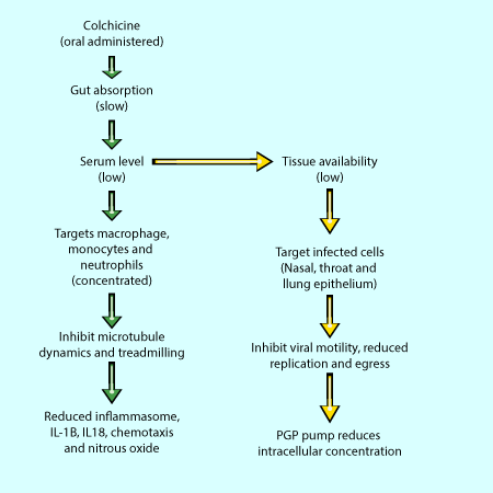
Considering CLN as an anti-inflammatory, the mechanism of action is thought to be multi-factorial acting through monocytes, macrophages and neutrophils. Documented effects include; reducing neutrophil chemotaxis7,8, inhibition of nitrous oxide production9, inhibiting the NACHT-LRRPYD containing protein 3 (NCPA3) inflammasome and downstream interleukin secretion10. In neutrophils, cell motility and chemotaxis are strongly inhibited because CLN is concentrated 13 to 68-fold inside these cells compared to other cell types11,21. This phenotype is thought to be a consequence of a lack of PGP-efflux pumps to remove the compound from the cytoplasm.

Above: Therapeutic pathways of colchicine through the body.

Above: Therapeutic pathways of colchicine through the body.

Colchicine Molecular Mechanisms
 
What are the molecular mechanisms that underlie CLN’s effects on migration, IL secretion, inflammasome and mitosis?  In short, CLN has two sites of action, the most well-known being the CLN binding site on ß-tubulin12,13, and the second is a site on the mitochondrial membrane which inhibits ATP from entering mitochondria via the voltage dependent channel14. Clearly the direct effect on tubulin and microtubules can to a large degree explain inhibition of motility, secretion, and mitosis due to microtubule’s critical role in those functions. Whereas the mitochondrial site may affect the amount of energy available for a neutrophil’s innate response, or the amount of acetyl-CoA for phospho-lipid synthesis in infected cells15 (see later).   
 
CLN’s therapeutic serum level is about 3 nM but varies widely between patients21. In vitro, CLN inhibits microtubule polymerization with an IC50 of 1-3 µM22, whereas its Kd (ratio of off rate versus on rate) is about 10-7M,23, which is almost impossible to measure due to its nearly 1-week dissociation half-life rate. The actual mechanism of inhibition in vitro was binding of a tubulin/colchicine complex to the ends of microtubules which inhibited the rates of polymerization and depolymerization by 50% at 0.16 µM24,25. The IC50 for dynamic instability of microtubules is closer to 100 nM26, which is closer to the 3 nM serum level when combined with the 13 to 67-fold increased concentration in neutrophils11,21. Therefore, the primary target of CLN in neutrophils could be microtubule dynamic instability and treadmilling.
 
Another interesting angle is the mitochondrial target of CLN14. Clearly, affecting mitochondrial function will reduce energy availability in the cell and decrease the rate of energy dependent processes such as polymer construction (RNA, fatty acids and protein synthesis). A connection between mitochondrial function and lipid metabolism is the acetyl-CoA shuttle27. Therefore, limiting mitochondrial function will reduce acetyl-CoA synthesis and result in less phospho-lipid production and could therefore reduce virion production28.
 
Colchicine Targets the Viral “Life-Cycle” 
 
From another perspective, the cytoskeleton is a key component of the viral “life-cycle” which includes infection, intra-cellular transport to a peri-nuclear position, replication, transport towards the plasma-membrane, and egress functions16,17,18,19. Within this scope, tubulin and microtubules are involved mostly in viral transport and replication, and Oliva et al. directly measured SARS-CoV-2 infection in vitro in the presence of CLN, among other cytoskeleton inhibitors, and found a 30% reduction (p<0.05 confidence with n=9 replicates) in viral spread20. A CLN binding site compound, podophyllotoxin, had even greater inhibition of viral infection and spread, a 60% reduction (p<0.0001), indicating that CLN site binding compounds can inhibit viral spread via interference with microtubule architecture in the absence of tissue and organism level inflammation. Other CLN site compounds such as mebendazole and febendazole also show efficacy, and have less side effects, which opens a path for therapy with less side effects than steroids or CLN.  However, much of the early work in 2020 utilized typical virus culturing cell types such as Vero E629, Caco-230 and A54931, which did not effectively replicate the characteristics of covid infected cells. Therefore, a pluripotent stem cell-derived AT2 cell line was developed by Hekman et al. 202032, which had improved alignment with alveoli AT2 cell type characteristics, and it resulted in a different pharmacological profile than the previous mentioned viral cell models.  
 
Colchicine Analogs: Potential Therapeutic for Covid-19 
 
Early and late stages Covid-19 therapy have quite different requirements, whereas early disease stage would require direct viral binding, replication and virion assembly inhibitors, the later stages require neutrophil and inflammation reduction. In vitro, CLN and its analogs appear to treat both aspects, however viral spread as judged by disease progression did not decrease significantly in clinical studies28, possibly due to CLN’s poor uptake rate and serum availability making for a poor effect11 (Fig.1). Other cytoskeletal components may be more efficacious targets e.g. F-actin and vimentin18 in the early stages of Covid-19.   
 
It is critical to note that late stage Covid-19 clinical trials of CLN were halted because it was no more efficacious over steroid use28 e.g. dexamethasone, which was being administered at the same time to the same patients receiving CLN. However, a mid-sized community trial (COLCORONA) administered to a pool of early PCR-positive infected people28 reported a 50% reduction in hospitalizations and deaths i.e, late-stage disease, but these were not significant in the small numbers collected. The early stage Covid-19 score was not affected by CLN.  
 
Neither pre-Covid 19 infection or late-stage CLN-alone trials have been attempted because steroids are quite effective at the late stage and CLN has a narrow therapeutic window in humans (0.5-2mg/day) to be administered with tolerable side effects. To improve the outcomes, second generation CLN-analogs and CLN binding site compounds are currently being assessed in the clinic. A recent example was a study with Sabizabulin from Veru Inc.33 The Phase II trial reported 82% reduction in mortality when 9 mg doses were given to moderate and high symptom patients.  As in the COLCORONA trial, there were low numbers of mortalities which made the data less substantial but still very encouraging, and even led to emergency use authorization for hospitalized moderate to severe COVID-19 patients at high risk for acute respiratory distress syndrome. Even so, based on the current information, it’s possible that different compounds will be needed for treating early and late Covid-19 cases as they manifest in different molecular models.

References

1. Kolle K, Martin MA, Antia R, Lopman B, and Dean NE. 2022. The changing epidemiology of SARS-CoV-2. Science, 375 (6585), p.1116-1121. DOI: 10.1126/science.abm4915
2. NIH webpage: https://www.covid19treatmentguidelines.nih.gov/therapies/immunomodulators/colchicine/
3.Ebers Papyrus, BC 1550. https://en.wikipedia.org/wiki/Ebers_Papyrus.
4. Schlesinger N, Firestein BL, Brunetti L. 2020. Colchicine in COVID-19: an Old Drug, New Use. Curr Pharmacol Rep, 6(4), p.137-145. doi: 10.1007/s40495-020-00225-6.
5. Montealegre-Gómez G, Garavito E, Gómez-López A, Rojas-Villarraga A, Parra-Medina R. 2021. Colchicine: A potential therapeutic tool against COVID-19. Experience of 5 patients. Reumatol Clin (Engl Ed), 17(7), p.371-375. doi: 10.1016/j.reumae.2020.05.008.
6. FDA webpage: https://clinicaltrials.gov/ct2/results?cond=covid-19&term=colchicine&cntry=&state=&city=&dist=
7. Borel JF, Feurer C. 1975. Chemotaxis of rabbit macrophages in vitro: inhibition by drugs. Experientia, 31(12). P.1437-9. doi: 10.1007/BF01923232. PMID: 814017
8. Paschke S, Weidner AF, Paust T, Marti O, Beil M, and Ben-Chetrit E. 2013. Technical advance: Inhibition of neutrophil chemotaxis by colchicine is modulated through viscoelastic properties of sub-cellular compartments. J. Leukoc. Biol. 94, p.1091-96.
9. Chia EW, Grainger R, and Harper JL. 2008. Colchicine suppresses neutrophil superoxide production in a murine model of gouty arthritis: a rationale for use of low dose colchicine. Br. J. Pharmacol. 153, 1288-95.
10. Martinon F, Petrilli V, Mayor A, Tardivel A, and Tschopp J. 2006. Gout-associated uric acid crystals activate the NALP3 inflammasome. Nature, 440(7081), p.237-241.
11. Fordham JN, Kirwan J, Cason J, and Currey HL. 1981. Prolonged reduction in polymorphonuclear adhesion following oral colchicine. Ann. Rheum. Dis. 40(6), p.605-608. Pubmed: 7332382.
12. Borisy GG, Taylor EW. 1967. The mechanism of action of colchicine. Binding of colchincine-3H to cellular protein. J. Cell Biol. 34(2):525-33. doi: 10.1083/jcb.34.2.525. PMID: 6068183
13. Downing K, Nogales E. 1998. Tubulin structure: insights into microtubule properties and functions. Current Opinion in Structural Biology, 8(6), p. 785-791. ISSN 0959-440X. doi.org/10.1016/S0959-440X(98)80099-7.
14. Amandine O. 2019. Tubulin-VDAC Interaction: Molecular Basis for Mitochondrial Dysfunction in Chemotherapy-Induced Peripheral Neuropathy. Frontiers in Physiology, 10, article 671, p.1-8. DOI:10.3389/fphys.2019.00671.
15. Shi L. and Tu BP. 2015. Acetyl-CoA and the Regulation of Metabolism: Mechanisms and Consequences. Curr Opin Cell Biol. 33, p. 125–131. doi:10.1016/j.ceb.2015.02.003.
16. Glebov OO. 2020. Understanding SARS-CoV-2 endocytosis for Covid-19 drug repurposing. FEBS Jo. 287, p. 3664-3671. Doi:10.1111/febs.15369.
17. Wen Z, Zhang Y, Lin Z, Shi K, Jiu Y. 2020. Cytoskeleton-a crucial key in host cell for coronavirus infection. J Mol Cell Biol. 12(12), p.968-979. doi: 10.1093/jmcb/mjaa042.
18. Kloc M, Uosef A, Wosik J, Kubiak JZ, Ghobrial RM. 2022. Virus interactions with the actin cytoskeleton-what we know and do not know about SARS-CoV-2. Arch Virol. 167(3), p.737-749. doi: 10.1007/s00705-022-05366-1.
19. Norris V, Ovádi J. 2021. Role of Multifunctional Cytoskeletal Filaments in Coronaviridae Infections: Therapeutic Opportunities for COVID-19 in a Nutshell. Cells. 10(7), p.1818. doi: 10.3390/cells10071818.
20. Oliva MÁ, Tosat-Bitrián C, Barrado-Gil L, Bonato F, Galindo I, Garaigorta U, Álvarez-Bernad B, París-Ogáyar R, Lucena-Agell D, Giménez-Abián JF, García-Dorival I, Urquiza J, Gastaminza P, Díaz JF, Palomo V, Alonso C. 2022. Effect of Clinically Used Microtubule Targeting Drugs on Viral Infection and Transport Function. Int J Mol Sci. 23(7), p.3448. doi: 10.3390/ijms23073448.
21. Chappey O, Niel E, Dervichian M, Wautier JL, Scherrmann JM, Cattan D. 1994. Colchicine concentration in leukocytes of patients with familial Mediterranean fever. Br J Clin Pharmacol. 38(1), p.87-9. doi: 10.1111/j.1365-2125.1994.tb04328.x.
22. Margolis RL, Wilson L. 1977. Addition of colchicine-tubulin complex to microtubule ends: the mechanism of substoichiometric colchicine poisoning. Proc Natl Acad Sci U S A. 74(8), p.3466-70. doi: 10.1073/pnas.74.8.3466.
23. Maekawa S. Dissociation constant of Tetrahymena tubulin-colchicine complex. J Biochem. 1978 Sep;84(3):641-6. doi: 10.1093/oxfordjournals.jbchem.a132169. PMID: 721798.
24. Bergen LG, and Borisy GG. 1983. Tubulin-colchicine complex inhibits microtubule elongation at both plus and minus ends. J Biol Chem. 258(7), p.4190-4. PMID: 6833250.
25. Skoufias DA, Wilson L. Mechanism of inhibition of microtubule polymerization by colchicine: inhibitory potencies of unliganded colchicine and tubulin-colchicine complexes. Biochemistry. 1992 Jan 28;31(3):738-46. doi: 10.1021/bi00118a015.
26. Vandecandelaere A, Martin SR, Schilstra MJ, Bayley PM. Effects of the tubulin-colchicine complex on microtubule dynamic instability. Biochemistry. 1994 Mar 15;33(10):2792-801. doi: 10.1021/bi00176a007.
27. Batchuluun B, Pinkosky SL, & Steinberg GR. 2022. Lipogenesis inhibitors: therapeutic opportunities and challenges. Nat Rev Drug Discov 21, 283–305. https://doi.org/10.1038/s41573-021-00367-2.
28. Tardif J-C, Bouabdallaoui N, L'Allier PL, Gaudet D, Shah B, Pillinger MH, et al. 2021. Colchicine for community-treated patients with COVID-19 (COLCORONA): a phase 3, randomised, double-blinded, adaptive, placebo-controlled, multicentre trial. The Lancet, 9(8), p.924-932. DOI:https://doi.org/10.1016/S2213-2600(21)00222-8 .
29. Bouhaddou M, Memon D, Meyer B, White KM, Rezelj VV, et al. 2020. The Global Phosphorylation Landscape of SARS-CoV-2 Infection. Cell, 182(3), p.:685-712.e19. doi: 10.1016/j.cell.2020.06.034.
30. Bojkova, D., Klann, K., Koch, B. et al. 2020. Proteomics of SARS-CoV-2-infected host cells reveals therapy targets. Nature, 583, p.469–472. https://doi.org/10.1038/s41586-020-2332-7.
31. Stukalov, A., Girault, V., Grass, V. et al. 2021. Multilevel proteomics reveals host perturbations by SARS-CoV-2 and SARS-CoV. Nature, 594, 246–252. https://doi.org/10.1038/s41586-021-03493-4.
32. Hekman RM, Hume AJ, Goel RK, Abo KM, Huang J, et al. 2020. Actionable Cytopathogenic Host Responses of Human Alveolar Type 2 Cells to SARS-CoV-2. Mol Cell. 80(6), p.1104-1122.e9. doi: 10.1016/j.molcel.2020.11.028.
33. Veru website: https://verupharma.com/pipeline/sabizabulin-for-covid-19/ .