1. Home
  2. Phosphotyrosine Detection Methods

Anti-Phosphotyrosine Validated Methods

Anti-Phosphotyrosine Validated Methods

Phosphotyrosine Overview

Tyrosine phosphorylation, a reversible process, is one of the most frequent post-translational modifications of proteins and is crucial in mediating signal transduction in eukaryotic cells after exposure to cytokines and growth factors (1). Anti-phosphotyrosine antibodies have been an important tools in studying the level of tyrosine phosphorylation of proteins in different cellular models. It has also played an important role in enriching phosphotyrosine peptides from trypsin-digested cell lysates. As a result, a large number of phosphopeptides have been identified under various physiological and pathological conditions with mass spectrometry technologies (2-3).

Cytoskeleton Image
Cytoskeleton Image
Cytoskeleton Image
Cytoskeleton Image

Phosphotyrosine Detection Protocols:

The following protocols provide optimized techniques for detecting proteins with a phosphotyrosine residue in Western blot, Immunoprecipitation, Immunfluorsecence, and ELISA techniques.

  • Western blot
  • Immunoprecipitation
  • Immunofluorescence
  • ELISA
  • Preperation of Pervanadate
Protocol 1 - Western blot using Phosphotyrosine Antibody

Reagents

Use as indicated in method at 1:500 dilution, sufficient for 100 ml of working strength Ab.

Method

  1. Run protein samples and control samples on SDS-PAGE.
  2. Equilibrate the gel in Western blot buffer (25 mM Tris pH 8.3, 192 mM glycine, 5% methanol) for 15 min at room temperature prior to electro-blotting.
  3. Transfer the protein to a PVDF membrane overnight at 40V at 4oC.
  4. Wash the membrane once with TBST for 10 minutes (10 mM Tris-HCl pH 8.0, 150 mM NaCl, 0.05% Tween 20).
  5. Block the membrane surface with 5% nonfat-dry milk in TBST for 60 min at room temperature with constant agitation.
  6. Incubate the membrane with a 1:500 dilution of anti-phosphotyrosine (Cat. # APY03) antibody diluted in TBST for 1h at room temperature or overnight at 4°C with constant agitation.
  7. Wash the membrane 3 times in TBST for 10 min each.
  8.  Incubate the membrane with an appropriate dilution (e.g., 1:20,000) of anti-mouse secondary antibody (e.g., goat anti-mouse HRP conjugated IgG from Jackson Labs., Cat. # 115-035-068) in TBST/3% non-fat milk for 30 min at room temperature.
  9. Wash the membrane 6 times in TBST for 10 min each.
  10. Use an enhanced chemiluminescence detection method to detect the signal (e.g., SuperSignal West Dura Extended Duration Substrate; ThermoFisher).

Results:

Western Blot Demonstration of APY03 Phosphotyrosine Specificity

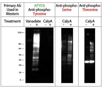
Legend: A431 cells were either treated (+) or untreated (-) with Calyculin A (CalyA: a serine/threonine phosphatase inhibitor, 50nM for 1 hour). NIH3T3 cells were either treated or untreated with H2O2-activated sodium orthovanadate (Vanadate: a specific tyrosine phosphatase inhibitor, 100 µM for 10 minutes). 16 µg of each lysate was resolved in SDS-PAGE and proteins were transferred to PVDF membrane. Membranes were probed as follows:

APY03 primary Ab: Detected tyrosine phosphorylated proteins in Vanadate-treated cells but not serine or threonine phosphorylated proteins in CalyA-treated cells.

Anti-phosphoserine & anti-phosphothreonine primary Ab: Detected serine/threonine phosphorylated proteins in CalyA-treated cells, demonstrating that phosphoserine/threonine proteins are present in CalyA treated lysates but are not detected by APY03. The anti-phosphoserine and anti-phosphothreonine Abs are from Millipore, Cat# AB1603 & AB1607 respectively.

Protocol 2 - Immunoprecipitation using Phosphotyrosine Antibody

Reagents

Use as indicated at 5 µl per IP reaction (200-500 µg total lysate per IP), sufficient for approximately 40 IP assays.

Method

  1. Add 5 µl of anti-phosphotyrosine (Cat. # APY03) to 500 µl of PBS pH 7.4 in a microfuge tube containing 30 µl of packed protein G agarose pre-equilibrated in PBS.
  2. Gently rotate the reaction for 1 hour at 4°C.
  3. Add 500 µl of PBST to the mixture and centrifuge for 1 minute at 4°C and 3000 rpm (960 x g). Addition of the PBST will prevent agarose from sticking to the microfuge tube walls.
  4. Discard supernatant and wash beads 3X in PBST (500 µl per wash).
  5. Add 200-500 µg of cell lysate (0.5 mg/ml protein in RIPA buffer) to the beads.
  6. Gently rotate the reaction at 4°C for 1 hour.
  7. Spin down agarose for 1 minute in 4°C at 3000 rpm (960 x g).
  8. Discard supernatant and wash beads with 800ul of wash buffer (50mM Tris pH7.5, 150mM NaCl and 1% IGEPAL). Spin down agarose for 1 minute in 4°C at 3000 rpm (960 x g).
  9. Repeat wash two more times. Carefully remove all supernatant without disturbing beads.
  10. Resuspend beads in 30 µl of 2X non reducing SDS sample buffer and incubate at room temperature for 5 minutes.
  11.  Spin down bead for 1 minute at 14,000 rpm (21,000 x g). Carefully remove 25 µl of supernatant and transfer to a new tube and add 1 µl of 2-mercaptoethanol. Boil for 5 minutes.
  12. Load samples on SDS-PAGE for subsequent western blot analysis. NOTE: to avoid detection of Ab heavy and light chains, we recommend using HRP conjugated CleanBlot (Thermo Scientific, #21230) as the secondary antibody reagent.

Results

IP of pervanadate-treated lysates from NIH3T3 cells

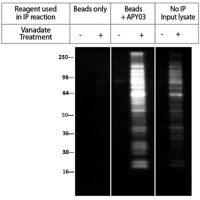
Legend: NIH3T3 cells were either treated (+) or untreated (-) with H2O2/orthovanadate (100 µM for 10min). Cell lysate was prepared in RIPA buffer and 200 µg of lysate per reaction was used for immunoprecipitation of tyrosine-phosphorylated proteins. APY03 was first bound to protein G beads and then incubated with cell lysate. For bead only control, cell lysate was incubated with protein G beads without APY03. Western blots of immunoprecipitated proteins were developed using APY03 at 1:500 dilution and CleanBlot (Thermo Scientific, #21230) as secondary antibody. No IP input lysate represents the signal from 5% of H2O2/orthovanadate treated or untreated NIH3T3 lysate. As shown in Figure 2, APY03 was able to enrich a wide range of tyrosine-phosphorylated proteins from NIH3T3 cells treated with H2O2-activated orthovanadate. No signal was detected with Protein G bead control without APY03.

Protocol 3 - Immunofluorescence using Phosphotyrosine Antibody (Cat. # APY03)

Reagents

Use as indicated below at 1:500-1:1000 dilution, sufficient for 100-200 ml of working strength Ab, approx. 1000-2000 IF slides.

Method

  1.  Plate cells to required density in suitable growth media on acid washed coverslips in tissue culture dish.
  2. Allow cells to grow for 24-48 hours then treat one set of coverslips with EGF (100ng/ml for 3 minutes, A431 cells), the other with H2O2-activated sodium orthovanadate (100 µM for 10 minutes, NIH3T3 cells).
  3. Fix cells by dipping coverslips in -20°C methanol for 5 minutes (it is convenient to use a porcelain coverslip rack for fixing and rinsing samples).
  4. Remove coverslips and dip into -20°C acetone for 2 minutes.
  5. Remove coverslips and air dry coverslips at room temperature.
  6. Rehydrate cells by dipping coverslips in PBS and then place coverslips, cell side up, on parafilm and apply 100-200 µl APY03 solution (1:500 in PBS for A431 cells and 1:1000 in PBS for NIH3T3 cells).
  7.  Incubate at room temperature for 60 minutes.
  8. Wash coverslips three times in PBS at room temperature over a 5 minute period.
  9.  Apply fluorescently labeled anti-mouse secondary antibody at manufacturers recommended dilution. For example, we use Alexa Fluor 555 goat anti-mouse (Life Technologies, Cat# A-21424) at 1:500 dilution in PBS.
  10.  Incubate at room temperature for 60 minutes.
  11.  Wash coverslips three times in PBS at room temperature over a 5 minute period.
  12. Rinse coverslips briefly in sterile water.
  13. Dip coverslips in absolute ethanol at room temperature for 2 minutes and transfer to blotting paper, cell side up, and allow to dry.
  14.  Place coverslips, cell side down, on glass slide with mounting media (e.g. EMS, Cat# 17987-10) and observe cells under fluorescence microscope.

Results

IF detection of phosphotyrosinated proteins in vanadate-treated cells

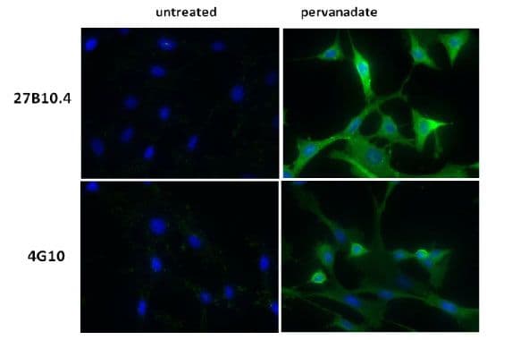
Legend: Human epidermoid carcinoma A431 cells, untreated (3A) or treated (3B) with EGF (100 ng/ml for 3 minutes), and NIH3T3, untreated (3C) or treated (3D) with H2O2-activated sodium orthovanadate (100 µM for 10 minutes), were stained as described in the method. Phosphotyrosine and nuclei were visualized in green fluorescence and blue DAPI staining, respectively.

Protocol 4 - ELISA using Phosphotyrosine Antibody (Cat. # APY03)

Reagents

Use as indicated below at 1:10,000 dilution, sufficient for approximately 40,000 assays.

Method

  1. Add 40 ng in 50 µl PBS of phosphopeptide BSA to the wells of ELISA plate.
  2. Incubated plate overnight at 4oC.
  3. Wash plate 2 times with 200 µl PBS.
  4. Add 200 µl of 3%milk in PBS and incubate at room temperature for 1 hour.
  5.  Wash plate 3 times with 200 µl PBS.
  6.  Add 50 µl of diluted APY03 (1:10,000 dilution in PBS). Incubated at room temperature for 1 hour with shaking.
  7. Wash plate with 3 times with 200 µl PBST.
  8. Add 50 µl of diluted goat anti-mouse secondary antibody (1:3000 dilution in 3%milk/PBST). Incubate at room temperature for 1 hour with shaking.
  9. Wash plate 3 times with 200 µl PBST.
  10. Add 50 µl of substrate to each well. Stop reaction by adding 50 µl of stop solution.
  11.  Measure OD at 490nm.

Results

ELISA assay demonstrating specificity of APY03 for phosphotyrosine

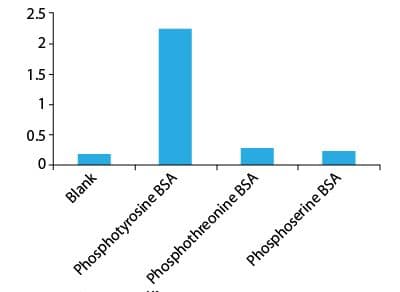
Legend: BSA was modified by conjugating with a library of phosphopeptides. 40ng of each phosphotyrosine, phosphothreonine and phosphoserine BSA were added to a individual well of a ELISA plate. APY03 at 1:10,000 dilution and goat anti mouse secondary antibody at 1:3000 were used to develop signal. Data shows that APY03 does not crossreact with phosphothreonine and phoophoserine modified BSA.

Protocol 5 - Preperation of Pervanadate

Sodium pervanadate should be made fresh and used immediately. Pervanadate is made by adding 6 µl of hydrogen peroxide to 44 µl of PBS in a microfuge tube. Add 50 µl of 200 mM sodium orthovanadate (the solution will turn yellow) and use immediately at the recommended dilutions.

Cytoskeleton Image

Useful Links

Overview of PTM Detection Techniques and Methods

SysPTM: A Systematic Resource for Post-Translational Modification

URL

http://lifecenter.sgst.cn/SysPTM/

What you can do

SysPTM provides a systematic and sophisticated platform for proteomic PTM research, equipped not only with a knowledge base of manually curated multi-type modification data, but also with four fully developed, in-depth data mining tools.

Literature

SysPTM: a systematic resource for proteomic research on post-translational modifications.

RegPhos: Regulatory Network in Protein Phosphorylation

URL

http://140.138.144.141/~RegPhos/index.php

What you can do

RegPhos is a resource to explore the protein kinase-substrate phosphorylation networks in human and mouse. 

Literature

RegPhos: a system to explore the protein kinase-substrate phosphorylation network in humans.

Related Products

  • Anti-Phosphotyrosine Monoclonal Antibody (Cat. # APY03)
  • Anti-Acetyl Lysine Monoclonal Antibody (Cat. # AAC03)
  • Anti-Ubiquitin Monoclonal Antibody (Cat. # AUB01)
  • More Antibodies

References:

  1. Machida, K. et al. (2003) Profiling the global tyrosine phosphorylation state. Mol. Cell. Proteomics 2, 215-233
  2. Blagoev, B. et al. (2004) Temporal analysis of phosphotyrosine-dependent signaling networks by quantitative proteomics. Nat. Biotechnol. 22, 1139-1145
  3. Schmelzle, K. et al. (2006) Temporal dynamics of tyrosine phosphorylation in insulin signaling. Diabetes 55, 2171-2179

Cytoskeleton, Inc
Contact UsMy Account

Tel: 303-322-2254

  • Blogs
  • FAQ's
  • Datasheets and MSDS
  • Protocols
  • Learning Documents
  • Videos
  • About Us
  • Careers
  • Distributors
  • All Services
  • Compound Screening
  • Protein Purification
  • Assay Development
  • Post-Translational Modification (PTM) Tools
  • Motor Protein Products
  • Live Cell Imaging Reagents
  • Actin Products
  • ECM Proteins
  • Small G-protein Products
  • Tubulin & FtsZ Products
  • General Protein Tools
Linkedin

© 2026 Cytoskeleton, Inc All Rights Reserved.

Privacy Policy

TEL: 303-322-2254

Log in into your Account
Cytoskeleton, IncCytoskeleton, Inc
Your quote is empty.
My Cart
( Items)
  • Cytoskeleton, Inc
  • Products
    • Featured New Product
    • Small G-protein Products
      • Small G-protein Activators & Inhibitors
      • Small G-protein Activation Assays
      • Small GTPase Affinity Beads
      • Small G-protein GEF/GAP and ELISA Kits
      • Small G-proteins
      • Modulator Proteins
      • Small G-protein Antibodies
      • Small G-protein Buffers
    • Post-Translational Modification (PTM) Products
      • Ubiquitination Tools
      • Acetyl-Lysine Tools
      • SUMOylation Tools
      • Phosphotyrosine Tools
      • Actin Oxidation Tools
      • Antibodies: PTMtrue™
      • Cell Lysis Tools
    • Tubulin & FtsZ Products
      • Tubulin Kits
      • Microtubules & Tubulin Proteins
      • Fluorescent and Labeled Tubulins
      • Microtubule-Associated Proteins (MAPs)
      • Live Cell Tubulin Reagents
      • Tubulin Antibodies
      • FtsZ Proteins
      • Tubulin Drugs and Buffers
    • Actin Products
      • Acti-stain™ Fluorescent Phalloidins
      • Actin Assays & Kits
      • Actin Proteins
      • Fluorescent and Labeled Actin
      • Actin Binding Proteins
      • Live Cell Actin Reagents
      • Actin Oxidation Tools
      • Actin Antibodies
      • Actin Drugs & Buffers
    • Live Cell Imaging Reagents
      • Live Cell Actin Reagents
      • Live Cell Tubulin Reagents
      • Small G-protein Activators & Inhibitors
      • Nuclear DNA Probes
      • Live Cell Membrane Probes
        • Plasma Membrane Probes
        • Membrane Tension Probes
        • Membrane Polarity Probes
      • Live Cell Mitochondrial Reagents
      • ECM Proteins
        • Fluorescent Fibronectin
        • Fluorescent Laminin
      • Live Cell Lipid Droplets
      • Other Organelles
      • Dyes/Substrates
    • ECM Proteins
      • Fluorescent Fibronectin
      • Fluorescent Laminin
    • Motor Protein Products
      • Motor Kits and Assays
      • Kinesin Proteins
      • Myosin Proteins
      • Dynein Proteins
      • Motor Protein Drugs & Buffers
    • General Protein Tools
      • Protein Quantitation
      • Cell Lysis Tools
      • ATPase & GTPase Assays
      • Other Protein Tools
    • GO-Blot V2 - Fully Automated And Programmable Western Blot Processor
  • Custom Services
    • All Services
    • Compound Screening
      • Tubulin Polymerization Assay Services
      • Kinesin ATPase Screening Services
      • Myosin ATPase Screening Services
      • GTP Exchange Factor (GEF) Assay Services
      • GTPase Activating Protein (GAP) Assay Services
      • FtsZ GTPase and Polymerization Assay Services
      • GTPase Activation Screening Services
    • Protein Purification
    • Assay Development
  • Resources
    • Resource Hub
      • Blogs
      • Videos
      • Protocols
      • Learning Documents
        • Ebook
    • FAQs
    • Datasheets and MSDS
  • About Us
    • About Us
    • Careers
    • Distributors
  • Contact Us
Your cart is empty.