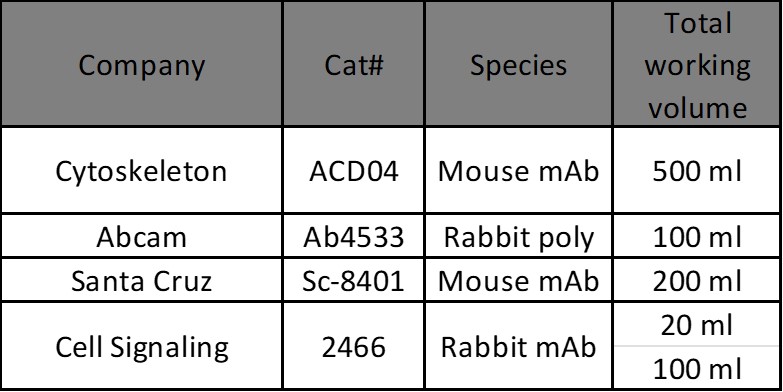
| Author | Title | Journal | Year | Article Link |
|---|
| Jayabal, Panneerselvam et al. | Roles of USP1 in Ewing sarcoma | Genes & Cancer | 2024 | PMID 38323120 |
| Ledoux, Benjamin et al. | Plasma membrane nanodeformations promote actin polymerization through CIP4/CDC42 recruitment and regulate type II IFN signaling | Science Advances | 2023 | ISSN 2375-2548 |
| Fu, Lisheng et al. | Septin11 promotes hepatocellular carcinoma cell motility by activating RhoA to regulate cytoskeleton and cell adhesion | Cell Death & Disease 2023 14:4 | 2023 | ISSN 2041--4889 |
| Serwe, Guillaume et al. | CNK2 promotes cancer cell motility by mediating ARF6 activation downstream of AXL signalling | Nature Communications 2023 14:1 | 2023 | ISSN 2041--1723 |
| Kamata, Tamihiro et al. | Statins mediate anti- and pro-tumourigenic functions by remodelling the tumour microenvironment | DMM Disease Models and Mechanisms | 2022 | ISSN 1754-8411 |
| Flores-Muñoz, Carolina et al. | The Long-Term Pannexin 1 Ablation Produces Structural and Functional Modifications in Hippocampal Neurons | Cells | 2022 | ISSN 2073-4409 |
| Chen, Bo et al. | Transient neuroinflammation following surgery contributes to long-lasting cognitive decline in elderly rats via dysfunction of synaptic NMDA receptor | Journal of Neuroinflammation | 2022 | ISSN 1742-2094 |
| Tyckaert, François et al. | Rac1, the actin cytoskeleton and microtubules are key players in clathrin-independent endophilin-A3-mediated endocytosis | Journal of cell science | 2022 | ISSN 1477-9137 |
| Kamata, Tamihiro et al. | Statins mediate anti- and pro-tumourigenic functions by remodelling the tumour microenvironment | DMM Disease Models and Mechanisms | 2022 | ISSN 1754-8411 |
| Li, Jingxuan et al. | Eva1a ameliorates atherosclerosis by promoting re-endothelialization of injured arteries via Rac1/Cdc42/Arpc1b | Cardiovascular Research | 2021 | ISSN 1755-3245 |
| Barbera, Stefano et al. | The C-type lectin CD93 controls endothelial cell migration via activation of the Rho family of small GTPases | Matrix Biology | 2021 | ISSN 1569-1802 |
| Jayabal, Panneerselvam et al. | NELL2-cdc42 signaling regulates BAF complexes and Ewing sarcoma cell growth | Cell Reports | 2021 | ISSN 2211-1247 |
| Howden, Jake D. et al. | α2β1 integrins spatially restrict Cdc42 activity to stabilise adherens junctions | BMC biology | 2021 | ISSN 1741-7007 |
| Hosseini, Kamran et al. | EMT-Induced Cell-Mechanical Changes Enhance Mitotic Rounding Strength | Advanced Science | 2020 | ISSN 2198-3844 |
| Gorisse, Laetitia et al. | Ubiquitination of the scaffold protein IQGAP1 diminishes its interaction with and activation of the Rho GTPase CDC42 | Journal of Biological Chemistry | 2020 | ISSN 1083-351X |
| Oni, Tobiloba E. et al. | SOAT1 promotes mevalonate pathway dependency in pancreatic cancer | Journal of Experimental Medicine | 2020 | ISSN 1540-9538 |
| Gu, Jiawen et al. | Rho-GEF trio regulates osteoclast differentiation and function by Rac1/Cdc42 | Experimental Cell Research | 2020 | ISSN 1090-2422 |
| Singh, Rajesh K. et al. | Dynamic Actin Reorganization and Vav/Cdc42-Dependent Actin Polymerization Promote Macrophage Aggregated LDL (Low-Density Lipoprotein) Uptake and Catabolism | Arteriosclerosis, Thrombosis, and Vascular Biology | 2019 | ISSN 1524-4636 |
| Huang, Yuxing et al. | Arp2/3-branched actin maintains an active pool of GTP-RhoA and controls RhoA abundance | Cells | 2019 | ISSN 2073-4409 |
| Dupraz, Sebastian et al. | RhoA Controls Axon Extension Independent of Specification in the Developing Brain | Current Biology | 2019 | ISSN 0960-9822 |
| Vestre, Katharina et al. | Rab6 regulates cell migration and invasion by recruiting Cdc42 and modulating its activity | Cellular and Molecular Life Sciences | 2019 | ISSN 1420-9071 |
| MacKeil, Jodi L. et al. | Phosphodiesterase 3B (PDE3B) antagonizes the anti-angiogenic actions of PKA in human and murine endothelial cells | Cellular Signalling | 2019 | ISSN 1873-3913 |
| Dudvarski Stanković, Nevenka et al. | EGFL7 enhances surface expression of integrin α 5 β 1 to promote angiogenesis in malignant brain tumors | EMBO Molecular Medicine | 2018 | ISSN 1757--4676 |
| Wu, Nan et al. | RCC2 over-expression in tumor cells alters apoptosis and drug sensitivity by regulating Rac1 activation | BMC Cancer | 2018 | ISSN 1471-2407 |
| Peretti, Amanda S. et al. | The R-Enantiomer of Ketorolac Delays Mammary Tumor Development in Mouse Mammary Tumor Virus-Polyoma Middle T Antigen (MMTV-PyMT) Mice | American Journal of Pathology | 2018 | ISSN 1525-2191 |
| Liu, Xiaolei et al. | Rasip1 controls lymphatic vessel lumen maintenance by regulating endothelial cell junctions | Development (Cambridge) | 2018 | ISSN 1477-9129 |
| Lam, Jonathan G.T. et al. | Host cell perforation by listeriolysin O (LLO) activates a Ca2+-dependent cPKC/Rac1/Arp2/3 signaling pathway that promotes Listeria monocytogenes internalization independently of membrane resealing | Molecular Biology of the Cell | 2018 | ISSN 1939-4586 |
| Li, Cao et al. | Regulation of staphylococcus aureus infection of macrophages by CD44, reactive oxygen species, and acid sphingomyelinase | Antioxidants and Redox Signaling | 2018 | ISSN 1557-7716 |
| Ghézali, Grégory et al. | Connexin 30 controls astroglial polarization during postnatal brain development | Development (Cambridge) | 2018 | ISSN 1477-9129 |
| Kawasaki, Natsumi et al. | TUFT1 interacts with RABGAP1 and regulates mTORC1 signaling | Cell Discovery | 2018 | ISSN 2056-5968 |
| Stypulkowski, Ewa et al. | The depalmitoylase APT1 directs the asymmetric partitioning of Notch and Wnt signaling during cell division | Science Signaling | 2018 | ISSN 1937-9145 |
| Vodicska, Barbara et al. | MISP regulates the IQGAP1/Cdc42 complex to collectively orchestrate spindle orientation and mitotic progression | Scientific Reports | 2018 | ISSN 2045-2322 |
| Bucka, Kenneth B. et al. | Local Arp2/3-dependent actin assembly modulates applied traction force during apCAM adhesion site maturation | Molecular Biology of the Cell | 2017 | ISSN 1939-4586 |
| Chang, Ting Ya et al. | Paxillin facilitates timely neurite initiation on soft-substrate environments by interacting with the endocytic machinery | eLife | 2017 | ISSN 2050-084X |
| Saito, Masaki et al. | Tctex‐1 controls ciliary resorption by regulating branched actin polymerization and endocytosis | EMBO reports | 2017 | ISSN 1469--221X |
| Takeuchi, Hiroki et al. | Intracellular periodontal pathogen exploits recycling pathway to exit from infected cells | Cellular Microbiology | 2016 | ISSN 1462-5822 |
| Seidelin, Jakob Benedict et al. | Cellular inhibitor of apoptosis protein 2 controls human colonic epithelial restitution, migration, and Rac1 activation | American Journal of Physiology - Gastrointestinal and Liver Physiology | 2015 | ISSN 1522-1547 |
| Thomas, Audrey et al. | Involvement of the Rac1-IRSp53-Wave2-Arp2/3 Signaling Pathway in HIV-1 Gag Particle Release in CD4 T Cells | Journal of Virology | 2015 | ISSN 0022--538X |
| Liu, Chunqiao et al. | Null and hypomorph Prickle1 alleles in mice phenocopy human Robinow syndrome and disrupt signaling downstream of Wnt5a | Biology Open | 2014 | ISSN 2046-6390 |
| David, Muriel D. et al. | The RhoGAP ARHGAP19 controls cytokinesis and chromosome segregation in T lymphocytes | Journal of Cell Science | 2014 | ISSN 0021-9533 |
| Valtcheva, Nadejda et al. | The orphan adhesion G protein-coupled receptor GPR97 regulates migration of lymphatic endothelial cells via the small GTPases RhoA and Cdc42 | Journal of Biological Chemistry | 2013 | ISSN 0021-9258 |
| Barrio, Laura et al. | TLR4 Signaling Shapes B Cell Dynamics via MyD88-Dependent Pathways and Rac GTPases | The Journal of Immunology | 2013 | ISSN 0022--1767 |
| Steele, Brian M. et al. | WNT-3a modulates platelet function by regulating small GTPase activity | FEBS Letters | 2012 | ISSN 0014-5793 |
| Mercer, Jason et al. | Vaccinia virus strains use distinct forms of macropinocytosis for host-cell entry | Proceedings of the National Academy of Sciences of the United States of America | 2010 | ISSN 0027-8424 |


