NOTE: This product has been discontinued for individual sale
Introduction
RhoA is one of most extensively studied member of Rho GTPase family. This family is involved in a wide range of cellular responses, including cytoskeletal reorganization, regulation of transcription, cell migration, cellular transformation and metastasis. RhoA is the predominant Rho isoform that is expressed in a large number of different cell types.
Product Uses Include
This antibody is recommended for detection of RhoA in human, mouse, rat or other extracts. The following protocols have been tested with this anti body:
- Western blot analysis: recommended
- Immunocytochemistry: recommended
RRID
AB_2884965
Material
The anti-RhoA antibody (Cat. # ARH05) is a mouse monoclonal antibody that only recognizes RhoA, not , RhoB, RhoC, Rac1, Rac2, Rac3, Cdc42 or H-Ras. Other commercially available RhoA antibodies recognize one or more of these close isoforms. ARH05 is supplied as a lyophilized white powder.
Figure 1 - Specificity and sensitivity of anti-RhoA by Western blot analysis.

Figure 1 legend: Recombinant small G-proteins and human platelet extract were separated by SDS-PAGE and transferred to a PVDF membrane according to the method given in this datasheet. Anti-RhoA was diluted 1 in 500 in TBST and western analysis was performed as detailed in the Western Blot Method section. Lane 1: 50 µg platelet extract, Lane 2: 10 µg platelet extract, Lane 3: 5 µg platelet extract, Lane 4: 10 ng His-RhoA, Lane 5: 5 ng His-RhoA, Lane 6: 2 ng His-RhoA, Lane 7: 100 ng His-RhoC. The recombinant and native RhoA band is visible at approximately 23 kD. No signal is visible with 100 ng of His-RhoC (Lane 7). The faint bands visible above His-RhoA represent a minor species of His-RhoA doublet. Exposure time 2 minutes. |
Figure 2: Polymerization of actin as measured by pyrene actin fluorescence
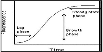
Table 1 - Reactivity of other commercially available RhoA antibodies
Supplier | RhoA | RhoB and RhoC | Rac sub-family | Ras sub-family | Cdc42 |
Cytoskeleton, Inc. (Cat. # ARH05) | Yes | No | No | No | No |
| Abcam (Cat. # ab54835) | Yes | Yes | No | No | No |
Santa Cruz (Cat. #SC-418) | Yes | NT | NT | NT | NT |
Upstate (Cat. # 05-778) | Yes | Yes | NT | NT | NT |
NT = Not tested
Other associated products:
Rho Inhibitor, cell permeable (Cat.# CT04)
Anti-Cdc42 monoclonal antibody (Cat.# ACD03)
Anti-Rac1 monoclonal antibody (Cat.# ARC03)
Cdc42 G-LISA™ Activation Assay (Cat.# BK127)
RhoA G-LISA™ Activation Assay (Cat.# BK124)
Rac1 G-LISA™ Activation Assay (Cat.# BK126)
Rac1,2,3 G-LISA™ Activation Assay (Cat.# BK125)
For product Datasheets and MSDSs please click on the PDF links below. For additional information, click on the FAQs tab above or contact our Technical Support department at tservice@cytoskeleton.com
Am. J. Physiol. Gastrointest. Liver Physiol. 2015; 308, G92-G99.
"Cellular inhibitor of apoptosis protein 2 controls human colonic epithelial restitution, migration, and Rac1 activation"
Author(s): Seidelin J.B. et al.
(See PubMed article)
Oncol Rep. 2015; 33, 1411-1417.
"Inhibition of microtubule-associated protein 1 light chain 3B via small-interfering RNA or 3-methyladenine impairs hypoxia-induced HO8910PM and HO8910 epithelial ovarian cancer cell migration and invasion and is associated with RhoA and alterations of the actin cytoskeleton"
Author(s): Tang Z. et al.
(See PubMed article)
Neurotoxicology. 2014; doi: 10.1016/j.neuro.2014.09.005.
"Amelioration of cisplatin-induced experimental peripheral neuropathy by a small molecule targeting p75NTR. "
Author(s): Friesland A. et al.
(See PubMed article)
J. Pharm. Pharmacol. 2014; 66, 418-427.
"Differential impact of the Bisphosphonate Alendronate on undifferentiated and terminally differentiated human myogenic cells."
Author(s): K. Shiomi et al.
(See PubMed article)
Mol. Cell Biol. 2013; 33, 2773-2786.
"Regulation of focal adhesion kinase activation, breast cancer cell motility, and amoeboid invasion by the RhoA guanine nucleotide exchange factor Net1. "
Author(s): Carr H.S., et al.,
(See PubMed article)
Int. J. Cancer. 2012; 130, 1273–1283.
"Human melanoma cells express FGFR/Src/Rho signaling that entails an adhesion-independent caveolin-1 membrane association. "
Author(s): Fecchi K., et al.
(See PubMed article)
PLoS ONE. 2012; 7: e38770.
"EGFR inhibition in glioma cells modulates Rho signaling to inhibit cell motility and invasion and cooperates with temozolomide to reduce cell growth. "
Author(s): Ramis G., et al.
(See PubMed article)
BMC Cancer. 2012; 12, 469.
"Vincristine enhances amoeboid-like motility via GEF-H1/RhoA/ROCK/Myosin light chain signaling in MKN45 cells. "
Author(s): Eitaki M., et al.
(See PubMed article)
Biomaterials. 2012; 33, 2902-2915.
"The RhoA-ROCK-PTEN pathway as a molecular switch for anchorage dependent cell behavior. "
Author(s): Yang S. and Kim H.-M.
(See PubMed article)
Question 1: What is the antigen that this antibody was raised against?
Answer 1: The RhoA specific antibody (Cat. # ARH05) is a mouse monoclonal IgM antibody and it was raised against amino acids 120-150 of the human RhoA protein.
Question 2: Is the IgM detected by normal anti-IgG HRP detection reagents?
Answer 2: Yes, there is high homology between IgG and IgM and hence most general anti-IgG detection reagents also detect IgM.
Question 3: What species has this antibody been tested against?
Answer 3: The RhoA specific antibody has been used to label RhoA proteins in human, canine, bovine, rat and mouse extracts.
If you have any questions concerning this product, please contact our Technical Service department at tservice@cytoskeleton.com.


