Rac1 G-LISA Activation Assay (Luminescence format) - 96 assays

G-LISA Rac 1 Activation Assay Biochem Kit (luminescence format)
$0.00

Product Uses Include

  • Rac1 signaling pathway studies
  • Rac1 activation assays with primary cells
  • Studies of Rac1 activators and inactivators
  • Rac1 activation assays with limited material
  • High throughput screens for Rac1 activation/inhibition

Introduction
The G-LISA series of Small G-Protein Activation Assays are ELISA based assays with which you can measure the GTP form of small G-proteins from lysates of cells or tissues and all in less than 3 h. The level of activation is measured with luminescence. For a more detailed introduction on G-LISA™ assays and a listing of other available G-LISA™ kits, see our main G-LISA™ page. The Rac1 G-LISA™ Activation Assay (Cat.# BK126) measures the level of GTP-loaded Rac1 protein in cell lysates, this is in contrast to Cat. BK125 which does not distinguish between activated Rac1,2 or 3. For a kit to measure RhoA activation please check webpage BK124.

The Rac1 G-LISA Activation Assay is very sensitive and has excellent accuracy for duplicate samples. See G-LISA™ FAQs tab on our G-LISA™ page for more details.

Kit contents
The kit contains sufficient reagents to perform 96 Rac activation assays. Since the Rac-GTP affinity wells are supplied as strips and the strips can be broken into smaller pieces, each kit can be used for anywhere from one to multiple assays. The following components are included in the kit:

  1. 96 Rac-GTP affinity wells (divisible into 96 individual wells)
  2. Lysis buffer
  3. Binding buffer
  4. Antigen presenting buffer
  5. Wash buffer
  6. Antibody dilution buffer
  7. Anti-Rac1 antibody
  8. HRP-labeled secondary antibody
  9. Positive control Rac1 protein
  10. Protease inhibitor cocktail (Cat. # PIC02)
  11. Luminescence detection reagents
  12. Precision Red™ Advanced protein assay reagent (Cat. # ADV02)
  13. Manual with detailed protocols and extensive troubleshooting guide

Equipment needed

  1. 96-well plate luminometer
  2. Multichannel or multidispensing pipettor
  3. Orbital microplate shaker capable of at least 200 rpm shaking (400 rpm is optimal)

Example results
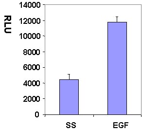
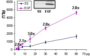
Serum starved Swiss 3T3 cells were stimulated with the Rac activating compound EGF and Rac activation was measured with the G-LISA method (Fig 1 and 2).
BK126_S-N

Figure 1. Rac1 activation by EGF measured by G-LISA™ kit BK126. Swiss 3T3 (mouse) cells were serum starved for 24 h and treated with EGF (10ng/ml for 2.5 min) or buffer only (SS). 10 µg of cell lysates were subjected to the G-LISA™ assay. Luminescence measured over 100 milli-second.
BK126_cv

Figure 2. Rac  activation by EGF measured by G-LISA™.  Swiss 3T3 cells were serum starved (SS) for 24 h and treated with EGF (10 ng/ml for 2 min). 60, 30, 15, 7.5, 2.5 µg of cell lysates were subjected to the G-LISA™ assay. Luminescence was measured over 10 milli-seconds. 500 µg of the same lysates were subjected to the traditional PAK pull-down assay (shown in inset, Cat.# BK035).

Go to main G-LISA™ page

G-LISA Products:
Cdc42 G-LISA™ Activation Assay, colorimetric format (Cat.# BK127)
Rac1,2,3 G-LISA™ Activation Assay, colorimetric format (Cat.# BK125)
RhoA G-LISA™ Activation Assay, colorimetric format (Cat.# BK124)
RhoA G-LISA™ Activation Assay, luminescence format (Cat.# BK121)

Associated Products:
Anti-Cdc42 monoclonal antibody (Cat.# ACD03)
Anti-Rac1 monoclonal antibody (Cat.# ARC03)
Anti-RhoA monoclonal antibody (Cat.# ARH03)

For product Datasheets and MSDSs please click on the PDF links below.   For additional information, click on the FAQs tab above or contact our Technical Support department at tservice@cytoskeleton.com

  •  G-LISA Activation Assay Technical Guide download here
  • For our G-LISA Data Analysis (Luminescence) Excel Template please download here.


    AuthorTitleJournalYearArticle Link
    He, Yi et al.CdGAP is a talin-binding protein and a target of TGF-β signaling that promotes HER2-positive breast cancer growth and metastasisCell reports2023
    Phuyal, Santosh et al.Mechanical strain stimulates COPII-dependent secretory trafficking via Rac1The EMBO Journal2022
    Nath, Anu S. et al.Modulation of the cell membrane lipid milieu by peroxisomal β-oxidation induces Rho1 signaling to trigger inflammatory responsesCell Reports2022
    Turgu, Busra et al.HACE1 blocks HIF1α accumulation under hypoxia in a RAC1 dependent mannerOncogene2021
    Garitano-Trojaola, Andoni et al.Actin cytoskeleton deregulation confers midostaurin resistance in FLT3-mutant acute myeloid leukemiaCommunications biology2021
    Zhu, Bili et al.Lipid oversupply induces CD36 sarcolemmal translocation via dual modulation of PKCζ and TBC1D1: An early event prior to insulin resistanceTheranostics2020
    Berthenet, Kevin et al.Failed Apoptosis Enhances Melanoma Cancer Cell AggressivenessCell Reports2020
    Crawford, Melissa et al.Essential Role for Integrin-Linked Kinase in Melanoblast Colonization of the SkinJournal of Investigative Dermatology2020
    Elshaer, Sally L. et al.Modulation of the p75 neurotrophin receptor using LM11A-31 prevents diabetes-induced retinal vascular permeability in mice via inhibition of inflammation and the RhoA kinase pathwayDiabetologia2019
    El-Naggar, Amal M. et al.HACE1 is a potential tumor suppressor in osteosarcomaCell Death and Disease2019
    Yan, Ting et al.Integrin αvβ3-associated DAAM1 is essential for collagen-induced invadopodia extension and cell haptotaxis in breast cancer cellsJournal of Biological Chemistry2018
    Kang, Jeong Hun et al.Protein kinase Cα as a therapeutic target in cancerProtein Kinase C: Emerging Roles and Therapeutic Potential2018
    Kai, Masahiro et al.Epigenetic silencing of diacylglycerol kinase gamma in colorectal cancerMolecular Carcinogenesis2017
    Jeong, Suk Yeong et al.Loss of Tpm4.1 leads to disruption of cell-cell adhesions and invasive behavior in breast epithelial cells via increased Rac1 signalingOncotarget2017
    Ruggiero, Carmen et al.Dosage-dependent regulation of VAV2 expression by steroidogenic factor-1 drives adrenocortical carcinoma cell invasionScience Signaling2017
    Kim, Jongshin et al.YAP/TAZ regulates sprouting angiogenesis and vascular barrier maturationJournal of Clinical Investigation2017
    Thuault, Sylvie et al.The RhoE/ROCK/ARHGAP25 signaling pathway controls cell invasion by inhibition of Rac activityMolecular Biology of the Cell2016
    Sylow, Lykke et al.Rac1 governs exercise-stimulated glucose uptake in skeletal muscle through regulation of GLUT4 translocation in miceJournal of Physiology2016
    Sarowar, Tasnuva et al.Enlarged dendritic spines and pronounced neophobia in mice lacking the PSD protein RICH2Molecular Brain2016
    Marchesin, Valentina et al.ARF6 promotes the formation of Rac1 and WAVE-dependent ventral F-Actin rosettes in breast cancer cells in response to epidermal growth factorPLoS ONE2015
    Reyes-Reyes, E. Merit et al.Mechanistic studies of anticancer aptamer AS1411 reveal a novel role for nucleolin in regulating Rac1 activationMolecular Oncology2015
    Yan, Yi et al.Augmented AMPK activity inhibits cell migration by phosphorylating the novel substrate Pdlim5Nature Communications2015
    Sylow, Lykke et al.Rac1 - a novel regulator of contraction-stimulated glucose uptake in skeletal muscleExperimental Physiology2014
    Radeva, Mariya Y. et al.PKA compartmentalization via AKAP220 and AKAP12 contributes to endothelial barrier regulationPLoS ONE2014
    Nakajima, Chikako et al.The lipoprotein receptor LRP1 modulates sphingosine-1-phosphate signaling and is essential for vascular developmentDevelopment (Cambridge)2014
    Kikuchi, K. et al.Protein kinase C iota as a therapeutic target in alveolar rhabdomyosarcomaOncogene2013
    Bray, Kristi et al.Cdc42 overexpression induces hyperbranching in the developing mammary gland by enhancing cell migrationBreast Cancer Research2013
    Sylow, Lykke et al.Rac1 signaling is required for insulin-stimulated glucose uptake and is dysregulated in insulin-resistant murine and human skeletal muscleDiabetes2013
    Redhu, Naresh Singh et al.Thymic stromal lymphopoietin induces migration in human airway smooth muscle cellsScientific Reports2013
    Eggers, Carrie M. et al.STE20-related kinase adaptor protein α (STRADα) regulates cell polarity and invasion through PAK1 signaling in LKB1-null cellsThe Journal of biological chemistry2012
    Sayedyahossein, Samar et al.Essential role of integrin-linked kinase in regulation of phagocytosis in keratinocytesFASEB Journal2012
    Vives, Virginie et al.The Rac1 exchange factor Dock5 is essential for bone resorption by osteoclasts2011
    Binker, Marcelo G. et al.TGF-β1 increases invasiveness of SW1990 cells through Rac1/ROS/NF-κB/IL-6/MMP-2Biochemical and biophysical research communications2011
    Kigel, Boaz et al.Plexin-A4 promotes tumor progression and tumor angiogenesis by enhancement of VEGF and bFGF signalingBlood2011
    McHenry, Peter R. et al.P190B RhoGAP has pro-tumorigenic functions during MMTV-Neu mammary tumorigenesis and metastasisBreast Cancer Research2010
    Johanna, Gutiérrez Vargas et al.Rac1 activity changes are associated with neuronal pathology and spatial memory long-term recovery after global cerebral ischemiaNeurochemistry international2010
    Kamai, Takao et al.Increased Rac1 activity and Pak1 overexpression are associated with lymphovascular invasion and lymph node metastasis of upper urinary tract cancerBMC Cancer2010
    Baumer, Y. et al.Role of Rac 1 and cAMP in endothelial barrier stabilization and thrombin-induced barrier breakdownJournal of Cellular Physiology2009
    Heckman-Stoddard, Brandy M. et al.Haploinsufficiency for p190B RhoGAP inhibits MMTV-Neu tumor progression2009
    Chastre, Eric et al.TRIP6, a novel molecular partner of the MAGI-1 scaffolding molecule, promotes invasivenessThe FASEB Journal2009
    Moniz, Sónia et al.WNK2 modulates MEK1 activity through the Rho GTPase pathwayCellular signalling2008
    Ramirez, Servio H. et al.Activation of Peroxisome Proliferator-Activated Receptor γ (PPARγ) Suppresses Rho GTPases in Human Brain Microvascular Endothelial Cells and Inhibits Adhesion and Transendothelial Migration of HIV-1 Infected MonocytesThe Journal of Immunology2008
    Petroll, W. Matthew et al.Dynamic assessment of fibroblast mechanical activity during rac-induced cell spreading in 3-D cultureJournal of Cellular Physiology2008
    Pontow, Suzanne et al.Antiviral Activity of a Rac GEF inhibitor Characterized with a Sensitive HIV/SIV Fusion AssayVirology2007

    Question 1:  There is less than a 2-fold difference in signal intensity between my positive control and lysis buffer blank.  Why?

    Answer 1:  To accurately measure luminescence signal intensity between the positive control and buffer blank, please check the instrument settings on the luminometer (see below for suggestions).  We also recommend running some “set-up” experiments with just the buffer blank and positive control to determine optimal settings for detecting the positive control signal 3-5 fold higher than the buffer blank.  It is also important to remember to use a fresh control protein tube for each run of positive control samples.  Do not store and re-use the positive control.

    Machine Settings

     Gain    

    Gain controls the sensitivity of the machine.  Most luminometers do not allow manual alteration of gain and use an auto-calibration or limited calibration function.  Turn off auto-settings and auto-calibration to use the machine in manual mode.  It is important to contact the luminometer manufacturer or consult the user’s manual to determine the best way to alter the machine sensitivity.  If gain can be altered, one should read at low, medium and high gains to determine the reading within the linear range of the assay (positive control should be 3-5X higher than blank). Gain range varies with instrument.  For example, gain in the Tecan GmbH SpectroFluor Plus ranges from 0 - 150 (where 150 is the highest).

     Integration Time

    This parameter can be varied on most machines.  It is a good idea to set the machine at the lowest integration time (usually 10 – 100 ms). Integration times greater than 200 ms are likely to read out of the linear range of the assay and may require lowering of gain or dilution of primary and/or secondary antibodies.

     Shaking

    Most machines give the shaking option.  The recommended setting is 5 sec shake, medium orbital speed before read.  This option is not essential to the assay.

     Temperature

    Room temperature

     Plate type

    Any setting that specifies 96 well flat, white will be sufficient.

     Filters

    Luminescence does not require excitation or emission filters so the filter spaces should be left blank.  If this is not an option, excitation can be set at any value and emission should be set between 400-500nm, with 430-445 as optimal setting.


    Question 2:  My arbitrary luminescence units (ALU) or relative luminescence units (RLU) are very different from what is depicted in the manual.  Why?

    Answer 2:  This is very typical as the luminescence units will vary from luminometer to luminometer based on the machine’s sensitivity and instrument settings.  The important information to take note of is what the relationship is between buffer blank and positive control luminescence values.  The positive control signal should be 3-5 fold greater than the buffer blank luminescence signal.  If that is the case, then the G-LISA assay is functioning in the linear range and experimental samples can now be processed.


     If you have any questions concerning this product, please contact our Technical Service department at tservice@cytoskeleton.com.