Spirochrome Probes for Bioimaging

Live-cell imaging (SiR and SIR700) probes developed by Spirochrome are cell-permeable compounds which stain microtubules (SiR-Tubulin), F-actin (SiR-Actin), Lysosomes (SiR-Lysosome) and chromosomal DNA (SiR-DNA) in living cells.  

Cytoskeleton is excited to introduce the next generation of Spirochrome’s live cell imaging probes - the SPY™ probes. SPY™ probes improve upon the SiR live cell imaging technology while also expanding the fluorophore labeling options for the study of F-actin, microtubules, and DNA in living cells.

Live cell F-actin imaging 2.0 with SPY650-FastAct™: This new probe is a unique fluorescent live cell actin probe that labels very dynamic actin filaments. Click Here to Learn More

Additional Spirochrome Probes and Tools:

Flipper-TR® is a live cell fluorescent membrane tension probe that simplifies the methodology for investigating changes in membrane tension when used in combination with standard fluorescence lifetime measurements. 

Benzylguanine substrates enables investigators to label their SNAP-tagged-fused-protein of interest with these fluorescent dyes; thus, harnessing the exceptional qualities of the SPY™ and SiR probes to enhance their investigation and understanding of their target protein. 

Click on the links below to learn more about these cutting-edge probes and tools for live cell imaging. 

Cytoskeleton, Inc. is the exclusive provider of Spirochrome, Ltd. products in North America.

Spirochrome technology is based on the proprietary fluorophore silicon rhodamine (SiR). SiR is a bright and photostable rhodamine-like dye. Its key features are its cell permeability, fluorogenic character and compatibility with super-resolution microscopy The fluorescence excitation and emission of SiR are in the far-red, reducing phototoxicity in live-cell imaging experiments. SiR is compatible with most microscopes as it can be used with standard Cy5 settings. The combination of all these properties set SiR-based probes apart from other fluorescent probes. Read more in this Nature Chemistry paper about the properties of SiR.


Fluorogenic probes for live-cell imaging of the cytoskeleton
SiR-actin and SiR-tubulin were recently introduced in a landmark paper published in Nature Methods. The probes combine minimal cytotoxicity with excellent brightness for fluorescence imaging of actin and tubulin. Combined with super-resolution microscopy, SiR-actin and SiR-tubulin permit live-cell imaging of the cytoskeleton with unprecedented resolution.


SiR-actin and SiR-tubulin can be used without transfection and without washing steps. An experiment that highlights these features is the use of SiR-actin to stain the actin fibers of erythrocytes by simply adding SiR-actin to whole blood. Furthermore, their far-red excitation and emission spectra make them compatible with genetically encoded reporters. 

References:

  1. Fluorogenic probes for live-cell imaging of the cytoskeleton, G. Lukinavičius et al., Nature Methods, 11, 731–733 (2014)
  2. A near-infrared fluorophore for live-cell super-resolution microscopy of cellular proteins G. Lukinavičius et al., Nature Chemistry, 5, 132–139 (2013).

Spirochrome's live cell probes have been cited hundreds of times over the past several years. SiR-Actin citations are described here. More individual product citations are listed on each product page.


AuthorTitleJournalYearArticle Link
Oh, Mijung et al.High extracellular glucose promotes cell motility by modulating cell deformability and contractility via the cAMP-RhoA-ROCK axis in human breast cancer cellsMolecular Biology of the Cell2023ISSN 1939-4586
Dang, David et al.Deep learning techniques and mathematical modeling allow 3D analysis of mitotic spindle dynamicsThe Journal of cell biology2023ISSN 1540-8140
Cui, Weiyingqi et al.Deciphering the Role of p60AmotL2 in Epithelial Extrusion and Cell DetachmentCells2023ISSN 2073-4409
Gutiérrez-Martínez, Enric et al.Actin-regulated Siglec-1 nanoclustering influences HIV-1 capture and virus-containing compartment formation in dendritic cellseLife2023ISSN 2050-084X
Subramani, Aravindh et al.Modulation of E-Cadherin Function through the AmotL2 Isoforms Promotes Ameboid Cell InvasionCells2023ISSN 2073-4409
Küster, Lukas et al.The actin bundling activity of ITPKA mainly accounts for its migration-promoting effect in lung cancer cellsBioscience Reports2023ISSN 1573-4935
Conway, James R.W. et al.Defined extracellular matrix compositions support stiffness-insensitive cell spreading and adhesion signalingProceedings of the National Academy of Sciences of the United States of America2023ISSN 1091-6490
Melica, Maria Elena et al.Preparation of Human Kidney Progenitor Cultures and Their Differentiation into PodocytesBio-protocol2023ISSN 2331-8325
Zheng, Minying et al.Cdc42 Regulates the Expression of Cytoskeleton and Microtubule Network Proteins to Promote Invasion and Metastasis of Progeny Cells Derived from CoCl2-induced Polyploid Giant Cancer CellsJournal of Cancer2023ISSN 1837-9664
Dunkley, Sam et al.Actin limits egg aneuploidies associated with female reproductive agingScience Advances2023ISSN 2375-2548
Deniz, Ilker A. et al.Mesenchymal stromal cell-associated migrasomes: a new source of chemoattractant for cells of hematopoietic originCell Communication and Signaling2023ISSN 1478-811X
Alvelid, Jonatan et al.Event-triggered STED imagingNature Methods 2022 19:102022ISSN 1548--7105
Wang, Li et al.Role of TRPC6 in periodontal tissue reconstruction mediated by appropriate stressStem Cell Research and Therapy2022ISSN 1757-6512
Caire, Robin et al.YAP promotes cell-autonomous immune responses to tackle intracellular Staphylococcus aureus in vitroNature Communications 2022 13:12022ISSN 2041--1723
Perez, Carolina Gomis et al.Rapid propagation of membrane tension at retinal bipolar neuron presynaptic terminalsScience Advances2022ISSN 2375-2548
Efremov, Yuri M. et al.3D nanomechanical mapping of subcellular and sub-nuclear structures of living cells by multi-harmonic AFM with long-tip microcantileversScientific Reports 2022 12:12022ISSN 2045--2322
Bauerly, Elisabeth et al.Impact of cilia-related genes on mitochondrial dynamics during Drosophila spermatogenesisDevelopmental Biology2022ISSN 0012--1606
Scherer, Katharina M. et al.A fluorescent reporter system enables spatiotemporal analysis of host cell modification during herpes simplex virus-1 replication2021PMID 33380421
Tsuchiya, Kenta et al.Ran-GTP Is Non-essential to Activate NuMA for Mitotic Spindle-Pole Focusing but Dynamically Polarizes HURP Near ChromosomesCurrent Biology2021ISSN 1879-0445
Kalargyrou, Aikaterini A et al.Nanotube‐like processes facilitate material transfer between photoreceptorsEMBO reports2021ISSN 1469--221X
Ordas, Laura et al.Mechanical Control of Cell Migration by the Metastasis Suppressor Tetraspanin CD82/KAI1Cells 2021, Vol. 10, Page 15452021ISSN 2073--4409
Kozawa, Kei et al.The CD44/COL17A1 pathway promotes the formation of multilayered, transformed epitheliaCurrent Biology2021ISSN 0960--9822
Fläschner, Gotthold et al.Rheology of rounded mammalian cells over continuous high-frequenciesNature Communications 2021 12:12021ISSN 2041--1723
Jin, Jianfeng et al.Pulsating fluid flow affects pre-osteoblast behavior and osteogenic differentiation through production of soluble factorsPhysiological Reports2021ISSN 2051--817X
Eggers, Nikolas et al.Cell-free genomics reveal intrinsic, cooperative and competitive determinants of chromatin interactionsNucleic Acids Research2021ISSN 0305--1048
Linklater, Erik S. et al.Rab40–cullin5 complex regulates eplin and actin cytoskeleton dynamics during cell migrationJournal of Cell Biology2021ISSN 1540-8140
Uhl, Bernd et al.uPA-PAI-1 heteromerization promotes breast cancer progression by attracting tumorigenic neutrophilsEMBO Molecular Medicine2021ISSN 1757--4684
Girardi, Francesco et al.TGFβ signaling curbs cell fusion and muscle regenerationNature Communications 2021 12:12021ISSN 2041--1723
Bain, Judith M. et al.Immune cells fold and damage fungal hyphaeProceedings of the National Academy of Sciences of the United States of America2021ISSN 1091-6490
Sala, Federico et al.Rapid Prototyping of 3D Biochips for Cell Motility Studies Using Two-Photon PolymerizationFrontiers in Bioengineering and Biotechnology2021ISSN 2296-4185
Bock, Fabian et al.Rac1 promotes kidney collecting duct integrity by limiting actomyosin activityJournal of Cell Biology2021ISSN 1540-8140
Titelbaum, Moran et al.Ezh2 harnesses the intranuclear actin cytoskeleton to remodel chromatin in differentiating Th cellsiScience2021ISSN 2589-0042
Salomaa, Siiri I. et al.SHANK3 conformation regulates direct actin binding and crosstalk with Rap1 signalingCurrent Biology2021ISSN 1879-0445
Eysert, Fanny et al.Alzheimer’s genetic risk factor FERMT2 (Kindlin-2) controls axonal growth and synaptic plasticity in an APP-dependent mannerMolecular Psychiatry2021ISSN 1476-5578
Raza, Sayyid et al.SOX9 is required for kidney fibrosis and activates NAV3 to drive renal myofibroblast functionScience Signaling2021ISSN 1937-9145
Vignaud, Timothée et al.Stress fibres are embedded in a contractile cortical networkNature Materials2021ISSN 1476-4660
Wallis, Samuel S. et al.The ESCRT machinery counteracts Nesprin-2G-mediated mechanical forces during nuclear envelope repairDevelopmental Cell2021ISSN 1878-1551
Monster, Jooske L. et al.An asymmetric junctional mechanoresponse coordinates mitotic rounding with epithelial integrityJournal of Cell Biology2021ISSN 1540-8140
Warmt, Enrico et al.Differences in cortical contractile properties between healthy epithelial and cancerous mesenchymal breast cellsNew Journal of Physics2021ISSN 1367-2630
Damenti, Martina et al.STED and parallelized RESOLFT optical nanoscopy of the tubular endoplasmic reticulum and its mitochondrial contacts in neuronal cellsNeurobiology of Disease2021ISSN 1095-953X
Choi, Yong Won et al.Senescent Tumor Cells Build a Cytokine Shield in Colorectal CancerAdvanced Science2021ISSN 2198-3844
Merlini, Mario et al.Microglial Gi-dependent dynamics regulate brain network hyperexcitabilityNature Neuroscience2021ISSN 1546-1726
Tabdanov, Erdem D. et al.Engineering T cells to enhance 3D migration through structurally and mechanically complex tumor microenvironmentsNature Communications2021ISSN 2041-1723
Jewett, Cayla E. et al.RAB19 Directs Cortical Remodeling and Membrane Growth for Primary CiliogenesisDevelopmental Cell2021ISSN 1878-1551
Wurzer, Hannah et al.Intrinsic Resistance of Chronic Lymphocytic Leukemia Cells to NK Cell-Mediated Lysis Can Be Overcome In Vitro by Pharmacological Inhibition of Cdc42-Induced Actin Cytoskeleton RemodelingFrontiers in Immunology2021ISSN 1664-3224
Morandell, Jasmin et al.Cul3 regulates cytoskeleton protein homeostasis and cell migration during a critical window of brain developmentNature Communications2021ISSN 2041-1723
Liu, Jiahao et al.Deep learning–enhanced fluorescence microscopy via degeneration decouplingOptics Express, Vol. 28, Issue 10, pp. 14859-148732020ISSN 1094--4087
Rafati, Yousef et al.Effect of microtubule resonant frequencies on neuronal cellshttps://doi.org/10.1117/12.25465692020Article Link
Logan, Gregory et al.Comparative analysis of taxol-derived fluorescent probes to assess microtubule networks in a complex live three-dimensional tissueCytoskeleton2020ISSN 1949--3592
Ueki, Hiroshi et al.Multicolor two-photon imaging of in vivo cellular pathophysiology upon influenza virus infection using the two-photon IMPRESSNature Protocols2020ISSN 1754--2189
Tamborino, Giulia et al.Cellular dosimetry of [177Lu]Lu-DOTA-[Tyr3]octreotate radionuclide therapy: the impact of modeling assumptions on the correlation with in vitro cytotoxicityEJNMMI Physics2020ISSN 2197-7364
Maraspini, Riccardo et al.Optimization of 2D and 3D cell culture to study membrane organization with STED microscopyJournal of Physics D: Applied Physics2020ISSN 1361-6463
Domart, Florelle et al.Correlating sted and synchrotron xrf nano-imaging unveils cosegregation of metals and cytoskeleton proteins in dendriteseLife2020ISSN 2050-084X
Kumari, Archana et al.Structural insights into actin filament recognition by commonly used cellular actin markersThe EMBO Journal2020ISSN 0261--4189
Schoenenberger, Angelina D. et al.Macromechanics and polycaprolactone fiber organization drive macrophage polarization and regulate inflammatory activation of tendon in vitro and in vivoBiomaterials2020ISSN 1878-5905
Stiff, Tom et al.Prophase-Specific Perinuclear Actin Coordinates Centrosome Separation and Positioning to Ensure Accurate Chromosome SegregationCell Reports2020ISSN 2211-1247
Valenzuela, José Ignacio et al.Localized Intercellular Transfer of Ephrin-As by Trans-endocytosis Enables Long-Term SignalingDevelopmental Cell2020ISSN 1878-1551
Reimer, Dorothea et al.B Cell Speed and B-FDC Contacts in Germinal Centers Determine Plasma Cell Output via Swiprosin-1/EFhd2Cell Reports2020ISSN 2211-1247
De Faveri, Francesca et al.LAP-like non-canonical autophagy and evolution of endocytic vacuoles in pancreatic acinar cellsAutophagy2020ISSN 1554-8635
Bouzhir, Latifa et al.Generation and quantitative characterization of functional and polarized biliary epithelial cystsJournal of Visualized Experiments2020ISSN 1940-087X
Pourcel, Lucille et al.Influence of cytoskeleton organization on recombinant protein expression by CHO cellsBiotechnology and Bioengineering2020ISSN 1097-0290
Takeuchi, Yasuto et al.Calcium Wave Promotes Cell ExtrusionCurrent Biology2020ISSN 0960-9822
Pal, Debadrita et al.Rac and Arp2/3-Nucleated Actin Networks Antagonize Rho During Mitotic and Meiotic CleavagesFrontiers in Cell and Developmental Biology2020ISSN 2296-634X
Roldán, Adrian Saul Jimenez et al.3,3′-Thiodipropanol As a Versatile Refractive Index-Matching Mounting Medium for Fluorescence MicroscopyBiomedical Optics Express2019ISSN 2156--7085
Maechler, Florian A. et al.Curvature-dependent constraints drive remodeling of epitheliaJournal of Cell Science2019ISSN 1477-9137
Hetmanski, Joseph H.R. et al.Membrane Tension Orchestrates Rear Retraction in Matrix-Directed Cell MigrationDevelopmental Cell2019ISSN 1878-1551
Janel, Sébastien et al.Stiffness tomography of eukaryotic intracellular compartments by atomic force microscopyNanoscale2019ISSN 2040-3372
Hu, Junyan et al.Distinct roles of two myosins in C. Elegans spermatid differentiationPLoS Biology2019ISSN 1545-7885
Tomba, Caterina et al.Laser-Assisted Strain Engineering of Thin Elastomer Films to Form Variable Wavy Substrates for Cell CultureSmall2019ISSN 1613-6829
Nagaraja, Surya et al.Histone Variant and Cell Context Determine H3K27M Reprogramming of the Enhancer Landscape and Oncogenic StateMolecular Cell2019ISSN 1097-4164
Tsygankova, Oxana M. et al.A unique role for clathrin light chain A in cell spreading and migrationJournal of Cell Science2019ISSN 1477-9137
Burger, Joyce et al.Fibulin-4 deficiency differentially affects cytoskeleton structure and dynamics as well as TGFβ signalingCellular Signalling2019ISSN 1873-3913
Balta, Emre et al.Spatial oxidation of L-plastin downmodulates actin-based functions of tumor cellsNature Communications2019ISSN 2041-1723
Vestre, Katharina et al.Rab6 regulates cell migration and invasion by recruiting Cdc42 and modulating its activityCellular and Molecular Life Sciences2019ISSN 1420-9071
Kahn, Olga I. et al.APC2 controls dendrite development by promoting microtubule dynamicsNature Communications2018ISSN 2041-1723
Neubert, Elsa et al.Chromatin swelling drives neutrophil extracellular trap releaseNature Communications2018ISSN 2041-1723
Schoenenberger, Angelina D. et al.Substrate fiber alignment mediates tendon cell response to inflammatory signalingActa Biomaterialia2018ISSN 1878-7568
Okumura, Masako et al.Dynein–dynactin–NuMA clusters generate cortical spindle-pulling forces as a multiarm ensembleeLife2018ISSN 2050-084X
Nozawa, Satoshi et al.Osteoblastic heparan sulfate regulates osteoprotegerin function and bone massJCI insight2018ISSN 2379-3708
Lickert, Sebastian et al.Morphometric analysis of spread platelets identifies integrin αiIbβ3-specific contractile phenotypeScientific Reports2018ISSN 2045-2322
Kschonsak, Yvonne T. et al.Activated ezrin controls MISP levels to ensure correct NuMA polarization and spindle orientationJournal of Cell Science2018ISSN 1477-9137
Zhao, Miao et al.Identification of the PAK4 interactome reveals PAK4 phosphorylation of N-WASP and promotion of Arp2/3-dependent actin polymerizationOncotarget2017ISSN 1949-2553
Petrini, Stefania et al.Aged induced pluripotent stem cell (iPSCs) as a new cellular model for studying premature agingAging2017ISSN 1945-4589
Magliocca, Valentina et al.Identifying the dynamics of actin and tubulin polymerization in iPSCs and in iPSC-derived neuronsOncotarget2017ISSN 1949-2553
Leite, Sérgio Carvalho et al.The Actin-Binding Protein α-Adducin Is Required for Maintaining Axon DiameterCell Reports2016ISSN 2211--1247
D'Este, Elisa et al.Subcortical cytoskeleton periodicity throughout the nervous systemScientific Reports2016ISSN 2045-2322
Lukinavičius, Gražvydas et al.Fluorogenic probes for live-cell imaging of the cytoskeletonNature Methods2014ISSN 1548-7105

Featured Papers

“Fluorogenic probes for live-cell imaging of the cytoskeleton”; G. Lukinavičius, L.Reymond, E. D’Este, A. Masharina, F. Göttfert, H. Ta, A. Güther, M. Fournier, S. Rizzo, H. Waldmann, C. Blaukopf, C. Sommer, D. W. Gerlich, H.-D. Arndt, S. W. Hell & K. Johnsson; Nature Methods 11, 731–733, 2014.


“STED Nanoscopy Reveals the Ubiquity of Subcortical Cytoskeleton Periodicity in Living Neurons”; E. D’Este, D. Kamin, F. Göttfert, A. El-Hady, S. W. Hell; Cell Reports , Volume 10 , Issue 8 , 1246 – 1251, 2015.


“A near-infrared fluorophore for live-cell super-resolution microscopy of cellular proteins”; G. Lukinavičius, K. Umezawa, N. Olivier, A. Honigmann, G. Yang, T. Plass, V. Mueller, L. Reymond, I. R. Corrêa Jr, Z. Luo, C. Schultz, E. A. Lemke, P. Heppenstall, C. Eggeling, S. Manley & K. Johnsson; Nature Chemistry 5, 132–139, 2013.


“Dynamic actin filaments control the mechanical behavior of the human red blood cell membrane”; D. S. Gokhin, R. B. Nowak, J. A. Khoory, A. de la Piedra, I. C. Ghiran and V. M. Fowler; Mol. Biol. Cell; February 25, 2015.


“A cleavable cytolysin-neuropeptide Y bioconjugate enables specific drug delivery and demonstrates intracellular mode of action”; V. M. Ahrens, K. B. Kostelnik, R. Rennert, D. Böhme, S. Kalkhof, D. Kosel, L. Weber, M. von Bergen and A. G. Beck-Sickinger; J. Control. Release; 209:170-178, 2015.


“Red Si–rhodamine drug conjugates enable imaging in GFP cells”; E. Kim, K. S. Yang, R. J. Giedt and R. Weissleder; Chem. Commun., 50, 4504-4507, 2014.


“A marginal band of microtubules transports and organizes mitochondria in retinal bipolar synaptic terminals”; M. Graffe, D. Zenisek, and J. Taraska; J. Gen Physiol. Vol. 146 No.1: 109-117, 2015.

Application Notes

“A Bright Dye for Live-Cell STED Microscopy”; S. Pitsch, I. Köster.

Q1. What is STED microscopy and how does it work?

A1. STED microscopy stands for Stimulated Emission Depletion microscopy.  It is one type of super resolution microscopy which allows the capture of images with a higher resolution than conventional light microscopy which is constrained by diffraction of light.  STED uses 2 laser pulses, one is the excitation pulse which excites the fluorophore, causing it to fluoresce.  The second pulse, referred to as the STED pulse, de-excites the fluorophore via stimulated emission in an area surrounding a central focal spot that is not de-excited and thus continues to fluoresce.  This is accomplished by focusing the STED pulse into a ring shape, a so-called donut, where the center focal spot is devoid of the STED laser pulse, conferring high resolution to the fluorescent area (Fig. 1; see Ref. 1 for more details on STED microscopy).

fig1-faq

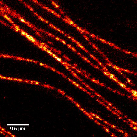
Figure 1. STED microscopic image of microtubules labeled with SiR-tubulin in human primary dermal fibroblasts.

Q2. Why is the SiR actin (or tubulin) probe good for STED microscopy?

A2. STED microscopy offers the ability to study cellular details on a nanometermolar scale in vivo.  To take advantage of this super resolution microscopy, one must be able to select with high specificity the area to be examined using fluorescent probes.  In addition, the fluorescent probes must be bright, photostable, exhibit no or little phototoxicity, be excited and emit in the far red spectrum.  In addition, if the probe is to be used for live cell imaging (thus avoiding fixation artifacts that occur when cells are fixed), high cell permeability is necessary.  The SiR actin and tubulin probes fulfill all of these requirements.  In short, the combination of STED and SiR probes allows for unparalleled fluorescent visualization of subcellular actin and tubulin/microtubule structures and their physical characterization in living cells, (see Fig. 2 and Ref. 2). 

Neuron_actin_1
Neuron_actin_closup2_1

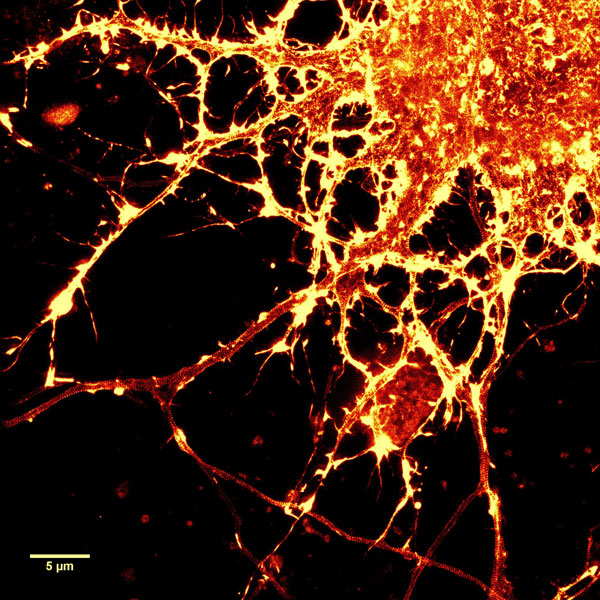
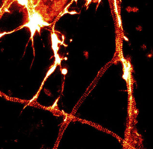
Figure 2. STED images of cultured rat hippocampal neurons stained with SiR-actin. Bottom image is a close-up view of part of the top image to clearly visualize actin rings (stripes) with 180 nm periodicity. Courtesy Of Elisa D'Este, MPI Biophysical Chemistry, Göttingen.


Q3. What are the filter sets for these probes?

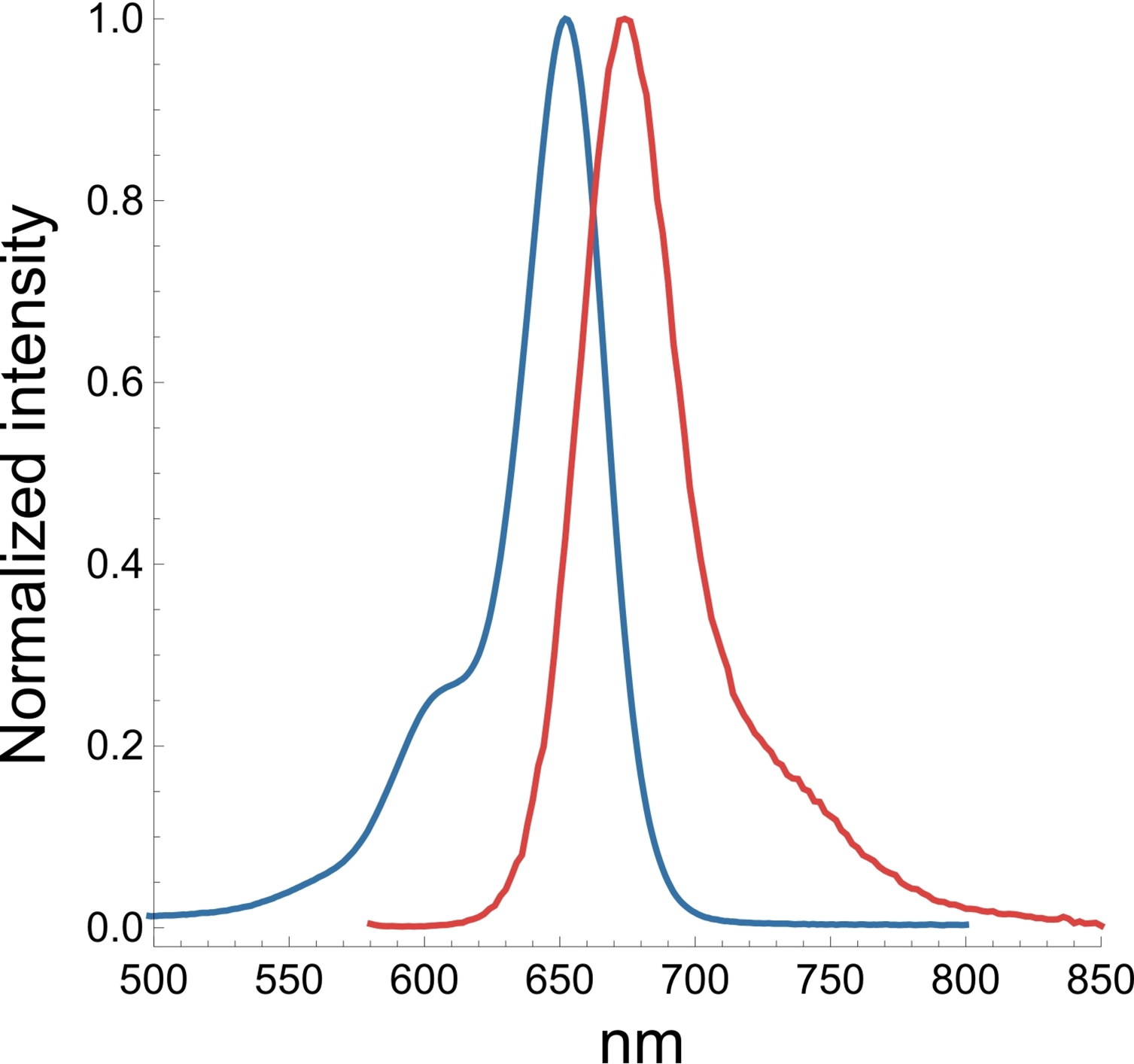
A3. The SiR actin and tubulin probes are visualized with standard Cy5 filters.  Optimal excitation is 650 nm and emission is 670 nm.  We recommend filters with an excitation of 630 + 20 nm and an emission of 680 + 20 nm (Fig. 3).  

faq-fig3

Q4. Why do the SiR probes have a low background compared to other fluorophores?

A4. SiR probes are excited by and emit light in the near infrared/far red spectral range, thus avoiding the use of shorter wavelengths such as blue and green light that typically autofluoresce, causing higher background signals.  SiR-coupled probes possess two physical states: 1. a non-fluorescent, closed off-state (spirolactone)  and 2. an open, highly fluorescent on-state (zwitterion).  The binding of the probe to its ligand target favors the highly fluorescent open state while the free unbound probe exists in the closed, non-fluorescent state (Fig. 4).  The fluorescence amplification is 100-fold from the unbound to bound state. This results in a highly sensitive biosensor in which the majority of fluorescence occurs only in the bound state (see Refs. 3 and 4). 

faq-fig4

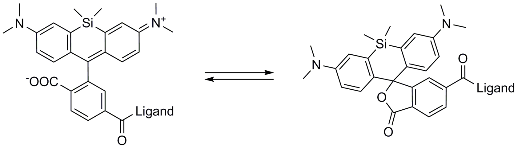
Figure 4. SiR derivatives exist in equilibrium between the fluorescent zwitterionic (open) form (left structure) and the non-fluorescent spiro (closed) form (right structure).


Q5: Are the SiR probes stable at room temperature?

A5: Yes, the probes are stable at room temperature for a few days.  However, it strongly depends on the probe and the solvent.  Thus, it is recommended to store all of the probes or solutions at –20°C.


Q6: Are SiR-actin and SiR-tubulin toxic to cells?

A6: Yes, above a certain threshold both probes show some effect on cell proliferation and altered actin or microtubule dynamics.  However, the probes are orders of magnitude less toxic than their parent drug.  In HeLa cells, neither actin nor microtubule dynamics were altered at concentrations below 100 nM.  At this concentration, SiR probes efficiently label microtubules and F-actin, allowing for the capture of high signal to noise images.


Q7: Do the probes work on fixed cells?

A7: SiR-actin probes can be used with PFA-fixed cells.  SiR-actin labels F-actin in PFA-fixed cells as efficiently as phalloidin derivatives.  SiR-tubulin labels microtubules only in ethyleneglycol-bis-succinimidyl-succinate (EGS)-fixed cells.  However, a selective labeling of centrosomal microtubules of PFA-fixed cells was observed.  SiR-actin and SiR-tubulin are not suitable for methanol-fixed cells.


Q8: Is it possible to image SiR-probes by STORM?

A8: No—under the very high light intensities typically used in STORM imaging, a phototoxic effect is observed on live cells.


Q9: Which organisms and tissues are stained by SiR-probes?

A9: This list describes only cell lines, tissues, or organisms that have been reported to work.  Omission of a cell line, tissue, or organism does not mean that the SiR-probes will not work with the specific cells, tissues, or organisms.


Homo sapiens: U2OS, fibroblasts, HeLa, HUVEC, MCF-10A, HCT-116, A549, erythrocytes

Mus musculus: C2C12, IA32, skeletal muscle, primary cardiomyocyte, primary oocyte

Rattus norvegicus: primary hippocampal neurons, primary cortical neurons, NRK

Cercopithecus aethiops: COS-7

Mesocricetus auratus: BHK

Drosophila melanogaster: Notum epithelium, S2

Didelphis marsupialis: OK cells


Q10. Do SiR-probes work in 3D cell cultures?

A10: Yes, the probes are able to stain cells in a 3D growth environment.


Q11: What are the correction factors CF260 and CF280 for the SiR fluorophore?

A11: CF260 = 0.116 and CF280  = 0.147


References

1. Hell S.W. and Wichmann J. 1994. Breaking the diffraction resolution limit by stimulated emission: stimulated-emission-depletion fluorescence microscopy. Opt. Lett. 19, 780-782.

2. D’Este E. et al. 2015. STED nanoscopy reveals the ubiquity of subcortical cytoskeleton periodicity in living neurons. Cell Rep. 10, 1246-1251.

3. Lukinavicius G. et al. 2013. A near-infrared fluorophore for live-cell super-resolution microscopy of cellular proteins. Nat. Chem. 5, 132-139.

4. Lukinavicius G. et al. 2014. Fluorogenic probes for live-cell imaging of the cytoskeleton. Nature Methods. 11, 731-733.

  1. ER Flipper-TR Kit for fluorescence cell membrane microscopy CY-SC021

    ER Flipper-TR Kit, a probe for measuring plasma-membrane tension.

    Learn More
  2. Flipper-TR Kit for fluorescence cell membrane microscopy CY-SC020
    Flipper-TR Kit, a probe for measuring plasma-membrane tension.
    Learn More
  3. Lyso Flipper-TR Kit for fluorescence cell membrane microscopy CY-SC022

    Lyso Flipper-TR Kit, a probe for measuring plasma-membrane tension.

    Learn More
  4. Mito Flipper-TR Kit for fluorescence cell membrane microscopy CY-SC023

    Mito Flipper-TR Kit, a probe for measuring plasma-membrane tension.

    Learn More